Article Text

Abstract
Background Following cued levodopa (LD) intake, endurance exercise showed a beneficial effect on scored motor performance in patients with Parkinson's disease (PD) in comparison with rest. This may result from an exercise induced increase in endogenous dopamine synthesis. As a result, beneficial effects on movement and reactivity may occur.
Objectives To measure reactivity and motor performance in a repeated fashion with instrumental tasks after cued administration of soluble 200 mg of LD/50 mg of benserazide.
Design PD patients consecutively performed paradigms, which assess reactivity and movement performance, after a standardised period of rest or of age-related, heart rate adapted endurance exercise on two consecutive days in a random order.
Results Reactivity and execution of simple and complex motion series were significantly better following exercise than after rest.
Discussion Endurance exercise has a beneficial effect on reactivity and movement behaviour in PD patients following cued application of LD probably due to an augmented synthesis and release of dopamine and other catecholamines and release in the prefrontal cortex, the nucleus accumbens and the basal ganglia. Small changes in catecholamine modulation of prefrontal cortex cells can have profound effects on the ability of the prefrontal cortex to guide behaviour. Previous exercise may also improve pedunculopontine nucleus function, which is involved in motor-related attention processes.
- Parkinson's disease
- levodopa
- exercise
- movement disorders
- neurophysiology
- motor
Statistics from Altmetric.com
Introduction
Treated patients with Parkinson's disease (PD) report a reduced exercise capacity due to a hypothetical limited effect of dopaminergic drugs or an increased need for them. Clinical trials excluded possible causes such as central dopamine deficiency, impaired gastrointestinal absorption and bioavailability of levodopa (LD), and mitochondrial muscle dysfunction. However, in PD patients, endurance exercise had a beneficial effect on non-blinded scored motor symptoms following cued LD intake compared with rest.1 However, even blinded rating is questionable, when comparing an exercise—with a rest condition due to onset of vegetative epiphenomena, such as sweating. This observed improved motor performance after endurance exercise may be due to release of endogenous dopamine, which contributes to regulation of reactivity and of movement.2 3 Conventional PD rating tools may not be sensitive and specific enough to capture these subtle changes in behaviour, which may be determined by simple instrumental tasks. Their execution depends on endogenous dopamine release in nigrostriatal and prefrontal brain structures in healthy humans, whereas an equal cued, dopaminergic stimulation is necessary to enable identical conditions in a cohort of PD patients to investigate whether endogenous dopamine release following exercise improves reaction- and movement behaviour.4 Various types of movement series exist. There are complex ones like peg insertion task. Its performance asks for additional involvement of prefrontal brain areas. They are similar to performed motion series of simple reaction time tasks, since they require execution of aimed movements. In contrast, tapping procedures are simple movement sequences, which are automatic and repetitive. They are more influenced by velocity with involvement of basal ganglia structures only. The objective of this exploratory, pilot trial was to assess reactivity and motion behaviour after cued dopaminergic stimulation and an interval of rest or of exercise in PD patients.
Subjects and methods
Subjects
Twenty-two treated idiopathic PD patients, diagnosed according to the UK Brain Bank criteria, (age: 61.05±2.08 (mean±SD) years; duration of PD: 4.21±0.61 years; UPDRS: 34.19±2.62; UPDRS I: 1.91±0.34; UPDRS II: 10.33±1.18; UPDRS III: 19.29±1.82; UPDRS IV: 2.57±0.62; HYS: 1.91±1.58; Mini Mental State Examination: 28.39±1.97) without unpredictable motor fluctuations were consecutively enrolled. PD patients, who had other medical conditions, which may affect the outcomes of the performed assessments, did not participate. The concomitant drug treatment consisted of LD/dopadecarboxylase inhibitor (N=22, 450±112.5 mg (mean±SD); entacapone N=9, 1000±200 mg; bromocriptine N=2, 30 mg each; cabergoline N=2, 2 mg, 4 mg; pergolide N=2, 2.25 mg, 8 mg; pramipexole N=11, ropinirole N=2, 9 mg, 15 mg; rotigotine N=1, 8 mg; rasagiline N=3, 1 mg each, selegiline N=3, 7.5 mg each; amantadine N=1, 300 mg).
Design
PD patients were put off their regular PD drug therapy for 12 h overnight. They had a standardised breakfast (300 kcal) in the morning at 07:00. Soluble LD/benserazide (200 mg/50 mg, Madopar LT) was applied after prior domperidone (Motilium, 40 mg) intake to avoid nausea 30 min later. A simple reaction-time paradigm (SRT) was performed 30 min before (point I) and 60, 90, 120 and 150 min after LD application (moments II–V). Conditions were identical except rest or exercise, both of which were performed in a random order (randomisation to condition sequence with sealed envelopes: Tanja Steiner) between moments I and II on two consecutive days in each participant (figure 1). The patient did not know in advance whether they would perform the exercise or the rest session on day 1, when performing the baseline investigation. One patient was tested at each session only. PD patients were trained with the use of the cycle ergometer on the day before start of the trial. The patients took their remaining drug regime after the study interval to reduce a bias of prior long-term dopamine substitution.
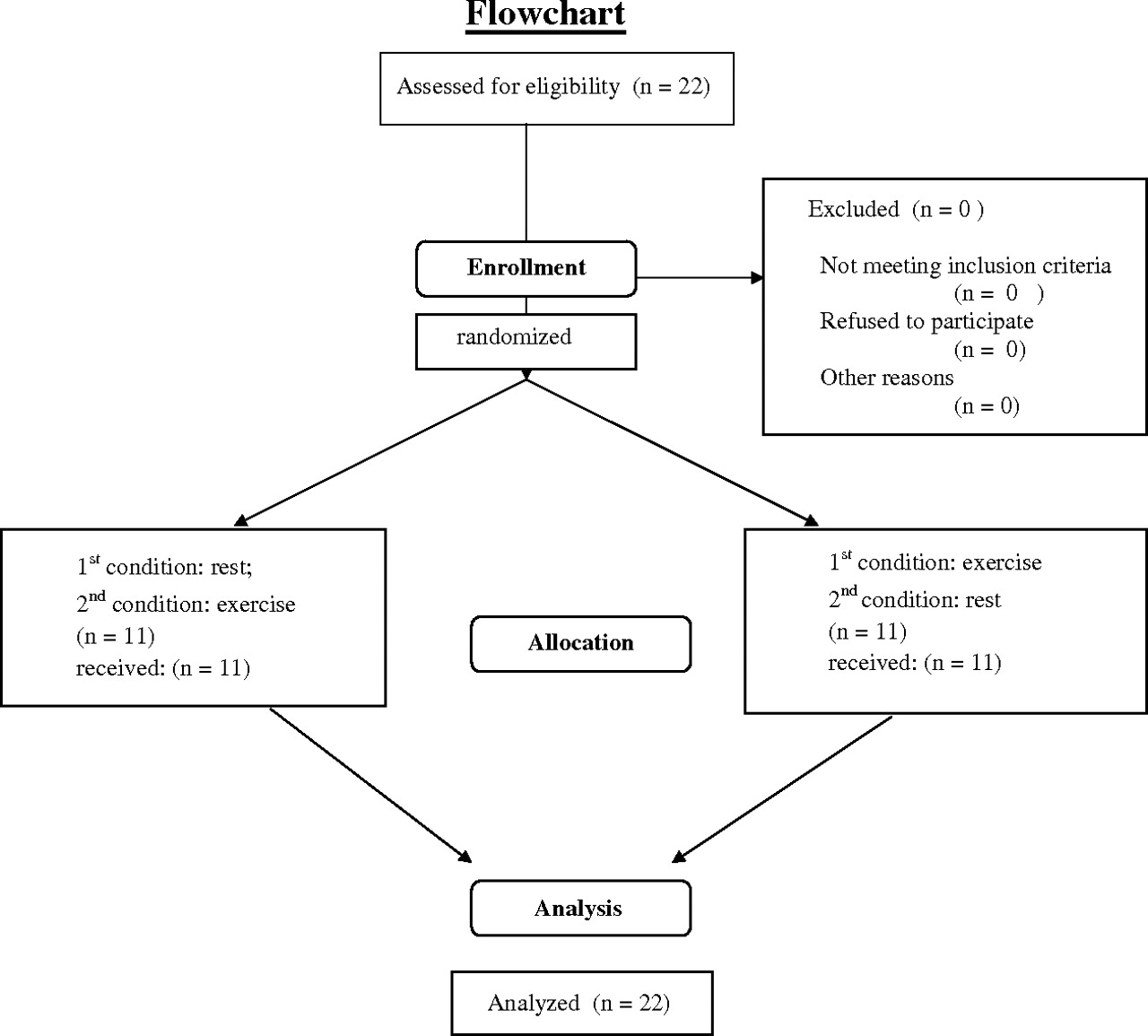
Study flow chart.
Exercise condition
Cycle ergometer exercise for the lower extremities was employed using a heart-rate adaptive training programme (Cateye Ergociser Modell EC-1600). By pedalling on the bike ergometer, the target heart rate was correlated with age. The heart frequency was measured by an ear-clip every 30 s and automatically relayed to the cycle workload. After reaching the age adjusted, required heart rate (formula: 200–age) in the 15 min lasting warm-up period, endurance exercise was performed with continuous, automatic adaptation of the bike pedal resistance over an hour lasting interval of exercise between moments I and II.
Rest session
The patients were in a lying position.
Simple reaction time paradigm (SRT)
The response to a visual SRT task (manufactured by Schuhfried, Mödling, Austria) was measured. The apparatus consisted of a 31 cm×42 cm rectangular surface with two stimulus lights (red and yellow), each coupled to the reaction button electrode 1 cm in diameter 15 cm equidistant from a central start button electrode. SRT performance did not depend on the red light, which was not presented and employed during the whole test procedure. The subject pressed the central start button with the index finger of the dominant hand (right: N=19; left: N=1; both hands: N=2). After the appearance of the yellow stimulus light, the subject had to switch off the light as quickly as possible by moving their finger from the central start button to the reaction button. Reaction time (RT) was considered as the elapsed interval between the onset of the yellow stimulus light and release of the start button. Movement time (MT) was the period between releasing the start button and pressing the reaction button. Thus, this paradigm only asks the subject to detect one stimulus and to produce the same response on every trial.5 Since the more affected hand of PD patients presents a slower SRT performance than the other less affected hand, PD patients were asked only to use the right dominant hand for this within-subjects comparison.6 RT and MT were assessed by a computer to millisecond accuracy. Twenty-eight RT trials were run. Out of 26 right answers, we used a truncated mean of values, which excludes measures greater than or less than 2 standard deviations (SD) of the mean value, for statistical analysis. In this trial, no participant fell within the 2 SD cut-off.
Peg insertion
In order to execute the peg insertion procedure (manufactured by Schuhfried) subjects were asked to transfer 25 pegs (diameter 2.5 mm, length 5 cm) from a rack into one of 25 holes (diameter 2.8 mm) in a computer-based contact board individually and as quickly as possible. The distance between rack and appropriate holes was exactly 32 cm. The board was positioned in the middle, and the task was carried out on each side. When transferring each peg from rack to hole, elbows were allowed to be in contact with the table. The interval between inserting of the first and the last pin initially with the right and then the left hand was assessed. Then, the data for the right and left hand were averaged to reduce the amount of data. The period for this task was measured by a computer in seconds with an accuracy below 100 ms.7
Tapping
In order to execute the tapping test (manufactured by Schuhfried), we instructed the individuals to tap as quickly as possible on a computer-based contact board (3 cm×3 cm) with a contact pencil for a period of 32 s after the initial flash of a yellow stimulus light. We did not control for peak height reached by the pencil. The board was positioned in the middle. When performing the task, elbows were allowed to be in contact with the table. We registered the number of contacts by means of a computerised device. We measured the tapping rate first with the right hand and then with the left.7
All participants were asked to familiarise themselves with the applied instrumental tests for an interval of 1 min to reduce or avoid learning and training effects on test performance on the day before.
Statistics
T-tests for comparisons between rest and exercise condition. ANCOVA (covariates: age, body weight, UPDRS) with a repeated measures design was employed for comparisons within one condition. The least significant difference t test was used for post-hoc comparisons. The level of significance was p<0.05. To minimise the effect of various baseline values, the differences (formula: value (moment I) minus value (II, III, IV, V each) while resting and during the exercise session were calculated.
Ethics
All participants gave written informed consent. The study was approved by the local ethics committee of the university.
Results
SRT
RT (F(4,84)=2.5, p<0.05; see figure 2A for post-hoc analysis) decreased after exercise and increased after rest (F(4,84)=2.55, p<0.05; figure 2B). There were no significant differences in baseline values (RT: exercise (vs) rest: p=0.1). Figure 2C compares the computed differences of baseline and each assessment point (formula see statistics section) between both conditions. Its outcomes support the observed RT reduction after exercise and the RT increase following rest.

Head: reaction time (RT) following exercise (A) and rest (B) at moments I–V, and (C) the computed differences of RT outcomes (I–II, I–III, I–IV, I–V) during the exercise—and the rest condition (bold, checkered). *p<0.05; **p<0.01; ***p<0.001 of the post-hoc test; I (baseline), II–V (after the intervention exercise or rest)=assessment moments.
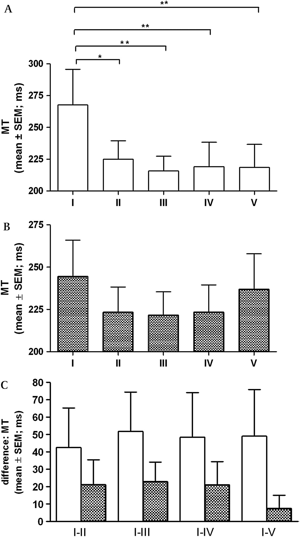
MT only significantly (exercise: F(4,84)=3.17, p<0.02 (figure 3A); rest: F(4,84)=1.86, n.s (figure 3B)) decreased following exercise. Baseline values between the rest and exercise session were not significantly different (MT: exercise vs rest (p=0.8)). Figure 3C shows the computed MT differences in baseline and each assessment point between both conditions. The number of correct answers did not change significantly after exercise (F(4,84)=0.12, NS) (data not shown), but decreased after rest (F(4,84)=6.89, p<0.001; post-hoc analysis: I (27.9±0.29; mean±SD) vs II (27.59±0.59) p<0.001; I vs III; ns.; I vs IV; ns.; I vs V ns).

Head: movement time (MT) after exercise (A) and rest (B) at moments I–V, and (C) the computed differences of MT outcomes (I–II, I–III, I–IV, I–V) during the exercise– and the rest condition (bold, checkered). *p<0.05; **p<0.01 of the post-hoc test; ***p<0.001 of the post-hoc test; I (baseline), II–V (after the intervention exercise or rest)=assessment moments.
Peg insertion
The interval for the peg insertion task decreased significantly after exercise (right: F(4,84)=16.04, p<0.001; figure 4A; left: F(4,84)=13.91, p<0.001; figure 4B) and resting (right: F(4,84)=4.90, p=0.0013; figure 4C; left: F(4,84)=6.85, p<0.001; figure 4D). Baseline values were not significantly different (peg insertion interval: exercise vs resting (right: p=0.16; left: p=0.38)). Figures 4E and 4F compare the computed differences of baseline and each assessment point between both conditions. The results demonstrate the significantly better improvement in peg insertion performance after exercise in particular with the right hand (figure 4E), but not with the left (figure 4F).

Head: interval for the peg insertion task after exercise ((A) right; (B) left hand) and rest ((C) right; (D) left hand) at moments I–V and computed differences (I–II, I–III, I–IV, I–V) during the exercise—and the rest condition ((E) right; (F) left hand) (bold, checkered). *p<0.05; **p<0.01; ***p<0.001 of the post-hoc test; I (baseline), II–V (after the intervention exercise or rest)=assessment moments.
Tapping
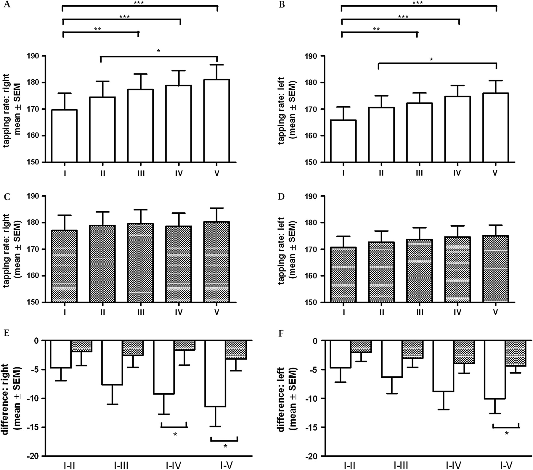
The tapping rate significantly increased after exercise (right: F(4,84)=5.70, p<0.001; figure 5A; left: F(4,84)=5.48, p<0.001; figure 5B), but not after rest (right: F(4,84)=0.73, ns; figure 5C; left: F(4,84)=2.48, ns; figure 5D). Baseline values did not differ significantly (right: p=0.35; left: p=0.46). Analogue to the previous analyses, significant differences between rest and exercise were found computed differences I–IV and I–V for the right hand figure 5E and the difference I–V for the left hand (figure 5F). This supports the finding of an improved tapping rate after exercise.

Head: tapping rate following exercise ((A) right; (B) left hand) and rest ((C) right; (D) left hand) during moments I–V respectively computed differences (I–II, I–III, I–IV, I–V) during the exercise—and the rest condition ((E) right; (F) left hand) (bold, checkered). *p<0.05; **p<0.01; ***p<0.001 of the post-hoc test; I (baseline), II–V (after the intervention exercise or rest)=assessment moments.
The covariates did not influence our results and no complaints of PD patients before and after exercise.
Discussion
Exercise and reactivity
The improved SRT performance following exercise may result from an augmented endogenous dopamine synthesis and release in various brain structures.5 Dopamine improves the nucleus accumbens function, which represents an interface between limbic and motor structures. The nucleus accumbens plays a major role in control of goal-directed actions and receives dopaminergic input from the ventral tegmental area. Behavioural studies consistently showed that the nucleus accumbens supports instrumental behaviours elicited by cues associated with drugs. Indirect stimulation of dopamine receptors in the nucleus accumbens by d-amphetamine shortened reaction times. Thus, stimulation of the nucleus accumbens with dopamine is involved in guiding the speed of instrumental responding.8 9 The impaired SRT execution after rest confirms previous outcomes on reactivity in PD patients with exact timing of both LD intake and test performance.10 An animal study also showed an impaired RT after cued dopaminergic stimulation in a particular species of fast-reacting rats.11 Sedative effects of LD may play a role, and release of fatigue-counteracting brain norepinephrine is lower during rest than during exercise.12 Therefore, the number of correct answers also decreased in the SRT paradigm after rest.
Exercise and motor performance
Execution of the peg insertion task was better after exercise than after rest in particular with the right dominant hand. This outcome confirms the prior observed behavioural pattern with the SRT paradigm with better test execution after exercise, and confirms that instrumental determination of disturbed movement performance is more valuable on the dominant side in PD patients.13 The peg insertion paradigm asks for execution of complex movement sequences with a complex interplay of an additional need for visual and spatial cognition, self-elaboration of internal strategies, sorting and planning. All these processes are influenced by the modulating role of striatal dopamine levels on association areas of the prefrontal cortex and the basal ganglia.4 14 Prior exercise supported endogenous dopamine synthesis and release in these brain areas.2 15 It is known that small changes in catecholamine modulation of prefrontal cortex cells can have profound effects on the ability of the prefrontal cortex to guide behaviour. We assume that these hypothetical higher prefrontal catecholamine levels following exercise supported the better peg insertion performance, as they also improved the execution of the attention-related components of this task.16 The tapping procedure only asks for performance of an automated, repetitive motion series without the need for attention- and concentration-related cognition load. Therefore, it depends more on dopamine-dependent basal ganglia function alone. Thus, tapping outcomes also improved due to an exercise-increased endogenous striatal dopamine release. Generally, one may also hypothesise that prior exercise additionally resulted in a better pedunculopontine nucleus function, which is involved in motor-related attention processes.17
Our study results also confirm findings that exercise provides benefit in PD patients in terms of overall motor function, in particular bradykinesia.18 This was also found with the improvement in UPDRS scores and in control with respect to coordination of grasping forces during the performance of a functional bimanual dexterity task. In particular, when PD patients were forced to exercise with a certain pedal rate and resistance 30% above their preferred voluntary one, a better motor performance and control were observed.19 In our trial, pedal rate and resistance was also continuously adapted to the heart rate. Therefore, we assume that the exercise condition within our trial was more similar to a more forced exercise paradigm and to an aerobic exercise condition.20 Forced exercise may lead to a shift in motor control strategy, from feedback to a greater reliance on feed-forward processes. This even suggests that forced exercise may alter central motor control processes.19 This is in line with trial outcomes, which describe dose-dependent benefits of exercise in early PD patients and suggest that even high-intensity exercise can normalise corticomotor excitability.21 Our results with a significantly better performance of instrumental motor tests following exercise support this hypothesis to a certain extent. We asked our participants to exercise above the voluntary level but not with a high intensity.
Limitations
Our explorative pilot trial did not investigate whether PD patients without any previous dopamine substitution also react different after a period of rest or exercise. Generally, test outcomes were better at baseline of the exercise condition. We cannot explain this phenomenon with dopamine-related reward- or expectation mechanisms related to exercise,22 since we performed a crossover design with 11 patients starting with exercise on day 1 and the remaining ones on day 2. However, all participants knew the test condition to be performed on the second day. This may reduce expectation and accordingly cause less release of endogenous dopamine and other catecholamines, since the participants were more adapted to the whole situation. Therefore, one may also hypothesise, that the more dopamine-related motor tests may be more sensitive to this hypothetical phenomenon than the reaction time paradigm. This was not the case, when the right- and left-handed motor tests were analysed separately, and accordingly no significant differences appeared concerning the SRT outcomes at baseline.
In conclusion, endurance exercise has a beneficial effect on reactivity and movement behaviour in PD patients following cued application of LD, probably due to an augmented synthesis and release of endogenous dopamine in various brain structures.
Acknowledgments
We thank T Steiner and C Stamm for technical assistance. We thank the participating patients.
References
Supplementary materials
Lay Summary
Files in this Data Supplement:
Footnotes
Competing interests None.
Patient consent Obtained.
Ethics approval Ethics approval was provided by the Bochum Ruhr University, Germany.
Provenance and peer review Not commissioned; externally peer reviewed.