Article Text

Abstract
Objective: To investigate the presence of serum anti-GT1a IgG in Guillain-Barré syndrome (GBS) and its relation to clinical manifestations.
Background: Several patients with GBS and bulbar palsy have been reported to have serum anti-GT1a IgG. Most, however, also have anti-GQ1b IgG. A previous study failed to detect GT1a in human cranial nerves, but GQ1b was abundant in human ocular motor nerves. Whether anti-GT1a IgG itself determines the clinical manifestations is not yet clear.
Methods: The association of clinical manifestations with the presence of anti-GT1a IgG and with its cross reactivity was investigated. An immunochemical study was performed to determine whether GT1a is present in human cranial nerves.
Results: Anti-GT1a and anti-GQ1b IgG were positive in 10% and 9% respectively of 220 consecutive patients with GBS. Patients with anti-GT1a IgG often had cranial nerve palsy (ophthalmoparesis, 57%; facial palsy, 57%; bulbar palsy, 70%), and 39% needed artificial ventilation. These features were also seen in patients with anti-GQ1b IgG. There was no significant difference between the two groups with respect to the frequency of clinical findings. An enzyme-linked immunosorbent assay showed that anti-GT1a IgG cross reacted with GQ1b in 75% of the patients, GD1a in 30%, GM1 in 20%, and GD1b in 20%. All five patients who carried anti-GT1a IgG that did not cross react with GQ1b had bulbar palsy, neck weakness, absence of sensory disturbance, and positive Campylobacter jejuni serology. Thin-layer chromatography with immunostaining showed that GT1a is present in human oculomotor and lower cranial nerves.
Conclusions: These findings provide further evidence that anti-GT1a IgG itself can determine clinical manifestations. The distinctive clinical features of patients with anti-GT1a IgG without anti-GQ1b activity distinguish a specific subgroup within GBS.
- Guillain-Barré syndrome
- anti-GT1a IgG
- cross reactivity
- Campylobacter jejuni
- GBS, Guillain-Barré syndrome
- MFS, Miller Fisher syndrome
- TLC, thin-layer chromatography
- ELISA, enzyme-linked immunosorbent assay
Statistics from Altmetric.com
- GBS, Guillain-Barré syndrome
- MFS, Miller Fisher syndrome
- TLC, thin-layer chromatography
- ELISA, enzyme-linked immunosorbent assay
Guillain-Barré syndrome (GBS) is a peripheral polyneuropathy characterised by acute onset of symmetrical muscle weakness with or without cranial nerve involvement. Some patients develop cranial nerve palsies early on, and generalised limb weakness appears as the illness progresses, whereas some experience only regional weakness throughout its course.1, 2 Several clinical variants have been proposed, cranial nerve involvement being a cardinal sign of some: ophthalmoparesis in Miller Fisher syndrome (MFS) or variant “sixth nerve paresis with paresthesia”,3, 4 blepharoptosis in variant “severe ptosis without ophthalmoplegia”,5 facial palsy in variant “facial diplegia and paresthesia”,4 and bulbar palsy in pharyngeal-cervical-brachial weakness or acute oropharyngeal palsy.5, 6
Mounting evidence indicates that the presence of serum antibody to ganglioside is closely related to clinical features. Anti-GQ1b IgG is specific to patients with MFS or GBS with ophthalmoplegia.7–9 Anti-GT1a IgG always coexists in these conditions, but its presence has been played down in the development of the disease because GQ1b is abundant in human oculomotor nerve, whereas GT1a is not detectable by biochemical analysis.8 Several patients have been reported with bulbar palsy and anti-GT1a IgG, but anti-GQ1b IgG has also been detected in most of them.6, 10–20 Whether anti-GT1a IgG itself determines the clinical manifestations or whether its activity contributes to the development of the disease has yet to be determined.
The first aim of this study was to determine the clinical features of anti-GT1a IgG positive patients with GBS and to compare them with those of anti-GQ1b IgG positive patients. The second was to examine GT1a ganglioside distribution in human cranial nerves in order to determine whether anti-GT1a IgG has a pathogenic role as an effector molecule in the development of cranial nerve palsies in GBS. Because anti-GT1a IgG that lacks cross reactivity with GQ1b was detected specifically in patients with the pharyngeal-cervical-brachial variant, we proposed that lack of antibody cross reactivity with GQ1b ganglioside is critical for the development of neurological features.21 The third aim was to clarify the range of cross reactivity of anti-GT1a IgG and to examine its relation to clinical features.
METHODS
Patients
Untreated serum samples were obtained within four weeks of the onset of neurological symptoms from 220 patients with GBS who had been referred to the neuroimmunological laboratory of Dokkyo University between August 1999 and August 2000 for anti-ganglioside antibody testing. None of them had been included in our previous studies.15, 21 The diagnosis of GBS was based on established clinical criteria.22 Patients who initially presented with ophthalmoplegia, ataxia, and areflexia and later developed generalised muscle weakness were also included with the GBS population. A questionnaire was used to collect the neurological data on the patients. Questions on the presence of consciousness disturbance, ophthalmoplegia, facial palsy, bulbar palsy, neck weakness, sensory disturbance, ataxia, dysautonomia, and arm or leg predominance limb weakness were completed by the primary physicians. For patients with anti-GT1a and/or anti-GQ1b IgG, we further reviewed their medical records to confirm that neurological deficits were invariably present and to obtain the results of electrophysiological studies and data on the functional disability grade. When the medical records did not contain adequate information, follow up faxes were sent to the primary physicians.
Serological assays
Serum IgG and IgM antibodies to GT1a, GQ1b, and other gangliosides (GM2, GM1, GM1b, GD1a, GalNAc-GD1a, GD1b, and GT1b) were measured by an enzyme-linked immunosorbent assay (ELISA) as described elsewhere.15 Serum was considered positive when antibody titre was 1000 or more. By this criterion, anti-GT1a IgG was positive in 11 (73%) of 15 patients with MFS, one (2%) of 50 patients with amyotrophic lateral sclerosis, and one (2%) of 50 healthy subjects.21 An absorption study, performed as described elsewhere,21 used microtitre plates coated with 10 pmol portions of ganglioside. Serum samples were added to the wells at a dilution that gave an absorbance of 1.0–2.0. After incubation at 4°C overnight, these samples were used as the primary antibodies in the standard ELISA. Absorption rate was expressed as percentage of absorbance obtained with and without absorption. The experiment was performed in triplicate, and the average decrease in anti-GT1a activity in three wells expressed as absorption rate. Evidence of recent Campylobacter jejuni infection was tested serologically as reported elsewhere.23
Thin-layer chromatography (TLC) with immunostaining
Total gangliosides were extracted from the cranial nerves and cauda equina of a patient as described elsewhere.24 The gangliosides were layered on TLC plates (Merck, Darmstadt, Germany) and then developed with chloroform/methanol/0.2% calcium chloride in water (50:45:10, by vol). The TLC plates were essentially immunostained as described elsewhere.17 In brief, plates were incubated with a patient's serum (diluted 1:100), which had high anti-GT1a (absorbance value, 0.339 at a dilution of 1:500 in the ELISA) and low anti-GQ1b (absorbance value, 0.023) IgG activities. This patient, reported on by Hashiguchi et al,11 experienced acute progression of dysphagia, dysarthria, neck weakness, and arm predominant limb weakness, but never exhibited ophthalmoplegia. After being washed, the plates were overlaid with peroxidase conjugated anti-(human γ chain specific) IgG (Dako, Glostrup, Denmark; 1:2000). After further washing of the plates, binding was made visible on x ray film by the use of ECL Western blotting reagents (Amersham International, Amersham, Buckinghamshire, UK).
Statistical analysis
Differences in proportions were examined by the χ2 or Fisher's exact test. Differences in medians were examined by the Mann-Whitney U test. A difference was considered significant at p<0.05. All statistical analyses were carried out with Statcel software (OMS, Saitama, Japan).
RESULTS
Antibodies to gangliosides
Anti-GT1a IgG was detected in 23 (10%) of the 220 patients and anti-GT1a IgM in two (1%). Patients with anti-GT1a IgG often had IgG antibodies against other gangliosides (GM1b (35%), GD1a (35%), GT1b (30%), GM1 (26%), GD1b (26%), GalNAc-GD1a (13%), GM2 (4%)), especially against GQ1b, 17 (74%) of these patients being positive for that antibody. Similarly, anti-GQ1b IgG was present in 19 (9%) of 220 patients and anti-GQ1b IgM in one (0.5%). Seventeen (89%) of the anti-GQ1b IgG positive patients also had anti-GT1a IgG.
Patients with anti-GT1a IgG often had cranial nerve palsy (ophthalmoplegia and/or diplopia, 57%; facial palsy, 57%; bulbar palsy, 70%), and 39% needed artificial ventilation (table 1). Serological evidence of recent C jejuni infection was found in 39%. These features, however, were as common as in patients with anti-GQ1b IgG. There were no significant differences between the clinical and laboratory findings for the anti-GT1a and anti-GQ1b IgG positive patient groups.
Clinical findings for patients with anti-GT1a or anti-GQ1b IgG
Absorption study
Twenty serum samples with anti-GT1a IgG were used. Anti-GT1a IgG activity decreased to less than 50% in all of them after incubation with GT1a, indicating that the absorption treatment was successful. Three samples were excluded because the antibody was not absorbed effectively by GT1a.
Anti-GT1a IgG was absorbed most often by GQ1b (fig 1), which absorbed it effectively (20% or more absorption rate) in 15 (75%) of the 20 patients' samples, and it was absorbed by GD1a in six (30%), GM1 in four (20%), and GD1b in four (20%). Although some patients who had anti-GT1a IgG lacking cross reactivity with GQ1b (closed circles in fig 1) had elevated antibody titres against some other gangliosides (table 2), their anti-GT1a IgG were absorbed only with GT1a ganglioside, which suggests that the antibody specifically recognises GT1a.
Clinical and laboratory findings for patients with anti-GT1a IgG lacking cross reactivity with GQ1b
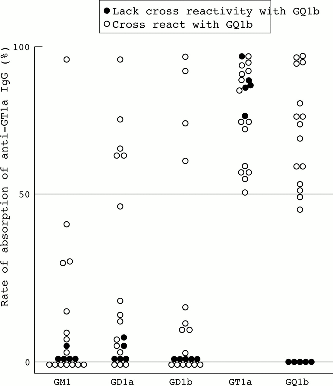
Plot of individual rates of absorption of anti-GT1a IgG. Five patients had anti-GT1a IgG lacking cross reactivity with GQ1b (clinical and laboratory features are given in table 2); 15 patients had anti-GT1a IgG with cross reactivity with GQ1b. Absorption rate was calculated from 1−((absorbance in well with serum with absorption treatment)/(absorbance in reference well with serum without absorption treatment)).
Fine specificity of anti-GT1a IgG and clinical findings
A statistical analysis was performed to examine the possible association between clinical findings and the cross reactivity of anti-GT1a IgG. Except for GQ1b, clinical manifestations were not associated with antibody cross reactivity with any ganglioside. Patients with anti-GT1a IgG lacking cross reactivity (less than 20% absorption rate) with GQ1b more often had a history of diarrhoea than did those with the reacting antibody (80% v 20%), and they had serological evidence of antecedent C jejuni infection (100% v 7%) (table 3). In contrast, a history of antecedent upper respiratory tract infection was rare in patients with the unreacting antibody (0% v 67%). These associations between cross reactivity and clinical features proved valid, even when the cut off values for judging “cross reactive” were defined as absorption rates of 5%, 10%, and 30%, rather than 20%.
Cross reactivity of anti-GT1a IgG with GQ1b
Five patients carrying anti-GT1a IgG with no cross reactivity with GQ1b had uniform manifestations and formed a distinct GBS subgroup (table 2); all had oropharyngeal and neck weakness, and serological evidence of recent C jejuni infection. None showed sensory loss. Only one (20%) had ophthalmoplegia, the condition characteristic of patients with anti-GT1a IgG. Statistical significance was not proven between these neurological features and antibody cross reactivity, probably because of the small patient population in this study.
TLC with immunostaining
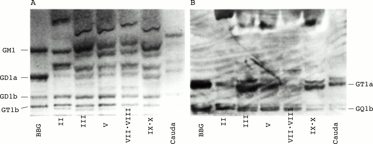
Immunostaining with a patient's serum that had anti-GT1a and anti-GQ1b IgG showed several bands in fractions from the oculomotor, glossopharyngeal, and vagal nerves, as well as from the bovine brain ganglioside mixtures, whereas none of these bands were visible in any of these cranial nerves when the stain was orcinol (fig 2). The mobility of the upper band(s) corresponded to GT1a and that of lower band(s) to GQ1b. This multiplicity of bands is thought to be due to differences in fatty acid composition. Interestingly, GQ1b was more weakly stained than GT1a in the glossopharyngeal and vagal nerve fraction, although it merely reflects that anti-GQ1b IgG activity was much lower than anti-GQ1b IgG of patient's serum used as the first antibody.
Immunostained thin-layer chromatograms. Plates stained with (A) orcinol/sulphuric acid for hexose, (B) serum IgG from a patient, with high anti-GT1a and low anti-GQ1b IgG activities. BBG (bovine brain ganglioside mixtures) and ganglioside fractions extracted from the IInd, IIIrd, Vth, VIIth/VIII, and IXth/X complex of cranial nerves or cauda equina were separated.
DISCUSSION
Ilyas et al25 reported that patients with anti-GT1a IgG tended to show dysphagia, but that the incidence of other cranial nerve palsies was not related to the presence of this antibody. Our much larger study showed that patients with anti-GT1a IgG often have multiple cranial nerve involvement, including ophthalmoplegia and facial as well as bulbar palsy. Anti-GT1a IgG rarely showed isolated elevated activity, often coexisting with anti-GQ1b IgG. We conclude that, on the whole, there is no difference in the clinical manifestations of the population of patients with anti-GT1a and anti-GQ1b IgG. Patients with GBS, who initially had low cranial nerve involvement, showed a spread to ocular motor nerve involvement in 33% and facial nerve palsy in 85%.2 Similarly, patients who initially showed oculomotor involvement often experienced the spread of weakness to the face and oropharynx. The common overlap of cranial nerve involvement in GBS is partially explained by the neurological manifestations of patients who have IgG antibodies that cross react with GT1a and GQ1b.
We previously showed that anti-GQ1b and anti-GM1 IgG have a broad spectrum of cross reactivity, which may account for the different clinical variations of GBS and MFS.26, 27 We here report that anti-GT1a IgG has various fine specificities, of which cross reactivity with GQ1b is the most common and related only to clinical and laboratory features, patients with anti-GT1a IgG that lacks cross reactivity with GQ1b being prone to develop oropharyngeal and neck weakness after C jejuni enteritis but rarely sensory deficit and ophthalmoparesis. These distinctive clinical features appear to characterise a specific subgroup within GBS. It should be noted that they are similar to the features of botulism, in which initially the cranial nerves are involved after diarrhoea and nausea, and then somatic paralysis without sensory loss occurs. Interestingly, gangliosides are the putative receptors of Clostridium botulinum toxins.28, 29 It has been hypothesised that Cl botulinum toxin and ganglioside antibodies bind to the same receptors, thereby causing muscle weakness in the region where the receptors are concentrated.30 When dry mouth, dizziness, and gastrointestinal symptoms are accompanied by neurological deficits, the condition can be differentiated from botulism by checking whether others who consumed the same food have a concurrent illness, by carrying out blood/stool assays for unbound toxin, as well as testing for antibodies to GT1a.
It is noteworthy that the anti-GT1a IgG that lacks cross reactivity with GQ1b also lacks cross reactivity with such other gangliosides as GM1, GD1a, and GD1b. This suggests that the detected antibody specifically recognised GT1a. There are two possibilities for the detection of anti-GQ1b IgG in the patients with anti-GT1a IgG lacking cross reactivity with GQ1b (patients 1 and 2 in table 2). We think that this is due to the much higher anti-GT1a activity than anti-GQ1b activity (128 000 v 4000, 128 000 v 8000). In these patients, very weak anti-GT1a activity (maybe only less than 1% of the total antibody) would cross react with GQ1b, resulting in the detection of anti-GQ1b IgG, whereas most anti-GT1a IgG does not cross react. The other possibility is that anti-GQ1b IgG that does not cross react with GT1a is present as well as anti-GT1a IgG that does not cross react with GQ1b.
We showed that GT1a ganglioside is present in human cranial nerves, including the glossopharyngeal and vagal ones. GT1a is a minor component of the human central and peripheral nervous systems.25, 31 A previous biochemical analysis failed to detect GT1a in the ganglioside fraction from human oculomotor nerve,8 whereas our immunochemical analysis detected it in various cranial nerves, including the oculomotor nerve. This discrepancy may reflect the fact that immunological methods are much more sensitive than chemical methods for detecting gangliosides. Another biochemical study showed a higher percentage of GQ1b ganglioside in the ocular motor and optic nerves than in the other cranial nerves, evidence that anti-GQ1b IgG functions in the pathogenic mechanisms of ophthalmoplegia in MFS and GBS.32 Because our GT1a composition analysis was not quantitative, the percentage of GT1a in each type of cranial nerve is not clear. Our data, however, support the possibility that anti-GT1a IgG has a pathogenic role as an effector molecule in the development of cranial nerve palsies in certain patients with GBS. Anti-GM1b IgG has also been reported to be common in patients with bulbar palsy.15, 17 Whether GM1b is a target antigen for serum antibody in GBS with cranial nerve disturbance must now be clarified.
We identified C jejuni as a possible inducer of anti-GT1a IgG that lacks cross reactivity with GQ1b in certain patients with GBS. C jejuni is a Gram-negative rod that has lipopolysaccharides with endotoxic activity. Lipopolysaccharides of a single C jejuni strain carry several core oligosaccharides, with terminal regions that mimic those of gangliosides.33 Some C jejuni strains isolated from patients with GBS or MFS have lipopolysaccharides bearing a GT1a-like structure.33–35 Whether the presence of a GT1a-like lipopolysaccharide is a factor that distinguishes the neuropathogenic from non-neuropathogenic strains is not clear. In this study, however, all five patients who had monospecific anti-GT1a IgG had serological evidence of previous C jejuni infection, evidence that the GT1a epitope of C jejuni lipopolysaccharides is an antigenic factor that induces the autoantibody and develops a neuropathy in some patients with GBS.
Acknowledgments
We thank Dr K Arimura (Kagoshima University, Kagoshima, Japan), Dr M Baba (Hirosaki University, Aomori, Japan), Dr M Mori (Chiba University, Chiba, Japan), Dr Y Mori (Daido Hospital, Aichi, Japan), Dr M Ogawa (Aomori Prefectural Central Hospital, Aomori, Japan), Dr B Okuda (Ehime Prefectural Central Hospital, Ehime, Japan) for providing the information on their patients. We also thank Ms Y Tsuchiya (Dokkyo University, Tochigi, Japan) for her technical assistance in testing the ganglioside antibody. M K is a Research Fellow (No 4734) of the Japan Society for the Promotion of Science. This research was supported in part by grants-in-aid from the Ryoichi Naito Foundation for Medical Research, grants-in-aid from the Uehara Memorial Foundation, grants-in-aid for Scientific Research (10780482 and 10557063 to N Y) from the Ministry of Education, Culture, Sports, Science and Technology of Japan, and a Research Grant for Neuroimmunological Diseases from the Ministry of Health and Welfare of Japan.