Article Text

Abstract
Background Chronic inflammatory demyelinating polyneuropathy (CIDP) is currently classified into ‘typical’ CIDP and ‘atypical’ subtypes such as multifocal acquired demyelinating sensory and motor neuropathy (MADSAM).
Objectives To assess the frequency of CIDP subtypes, and to elucidate clinical and electrophysiological features, and treatment response in each subtype.
Methods We reviewed data from 100 consecutive patients fulfilling criteria for CIDP proposed by the European Federation of Neurological Societies and the Peripheral Nerve Society. The Kaplan-Meier curve was used to estimate long-term outcome.
Results Patients were classified as having typical CIDP (60%), MADSAM (34%), demyelinating acquired distal symmetric neuropathy (8%) or pure sensory CIDP (1%). Compared with patients with MADSAM, patients with typical CIDP showed more rapid progression and severe disability, and demyelination predominant in the distal nerve segments. MADSAM was characterised by multifocal demyelination in the nerve trunks. Abnormal median-normal sural sensory responses were more frequently found for typical CIDP (53% vs 13%). Patients with typical CIDP invariably responded to corticosteroids, immunoglobulin or plasmapheresis, whereas patients with MADSAM were more refractory to these treatments. The Kaplan-Meier analyses showed that 64% of patients with typical CIDP and 41% of patients with MADSAM had a clinical remission 5 years later (p=0.02).
Conclusions Among the CIDP spectrum, typical CIDP and MADSAM are the major subtypes, and their pathophysiology appears to be distinct. In typical CIDP, the distal nerve terminals and possibly the nerve roots, where the blood–nerve barrier is anatomically deficient, are preferentially affected, raising the possibility of antibody-mediated demyelination, whereas cellular immunity with breakdown of the barrier may be important in MADSAM neuropathy.
- EMG
- NEUROPHYSIOL, CLINICAL
- NEUROIMMUNOLOGY
Statistics from Altmetric.com
Introduction
Chronic inflammatory demyelinating polyneuropathy (CIDP) is an immune-mediated neuropathy with a heterogeneous clinical manifestation.1–3 CIDP should be considered in any patients with progressive symmetric or asymmetric demyelinating polyneuropathy, because it is a treatable disease. Randomised controlled trials have indicated that approximately two-thirds of patients with CIDP improve by treatment with corticosteroids, immunoglobulin and plasmapheresis.4 ,5 However, up to 21% of patients with CIDP are refractory to these immune therapies and have severe disability.6–8 The different response to immune treatments may depend on the different pathophysiology in each CIDP subtype, and some factors could determine treatment response and outcomes.
The concept of CIDP has changed somewhat over the past two decades. The heterogeneity partly results from a different definition of the disorder. Classical CIDP is clinically defined as symmetric polyneuropathy involving the proximal and distal muscles, including a relatively uniform group of patients.9 Research criteria proposed by the American Academy of Neurology (AAN) defined a clinical inclusion criterion as “motor or sensory dysfunction of more than one limb”.10 According to the AAN criteria, a number of variants such as multifocal acquired demyelinating sensory and motor neuropathy (MADSAM or Lewis-Sumner syndrome), multifocal motor neuropathy, and distal acquired demyelinating symmetric (DADS) polyneuropathy could be included in CIDP. 11 ,12
In 2005, the Joint Task Force of the European Federation of Neurological Societies and the Peripheral Nerve Society (EFNS/PNS) classified CIDP into clinical subtypes. In this guideline (updated in 2010),13 classical CIDP is categorised as ‘typical CIDP’, and ‘atypical CIDP’ includes MADSAM/asymmetric CIDP, DADS, and pure motor or sensory CIDP. These conditions are still considered CIDP but have different features, whereas in EFNS/PNS criteria, multifocal motor neuropathy and antimyelin-associated glycoprotein (anti-MAG) neuropathy were excluded from CIDP, mainly because of different treatment responses. All the subtypes of CIDP share common features, and acquired peripheral nerve demyelination presumably mediated by autoimmune responses, but the different phenotypes suggest a different pathophysiology and immunopathogenesis.
Electrophysiological examination can provide important information about the distribution of demyelinative lesions along the nerves, and it is proposed that distribution patterns of demyelination may determine the clinical phenotypes.14 ,15 The aim of this study is to provide the frequency of each subtype of CIDP in a consecutive patient cohort, and to elucidate differences in electrophysiological profiles, response to treatments, long-term prognosis, and thereby in the pathophysiology among the CIDP subtypes.
Methods
Patients
Between 1994 and 2013, 139 patients with chronic demyelinating neuropathy and features suggesting CIDP were seen at Chiba University Hospital, Chiba, Japan (figure 1). Of these, 23 patients had multifocal motor neuropathy, and 8 suffered demyelinating neuropathy with anti-MAG. Eight patients had a coincidental other neuroimmunological disorder or cerebral palsy. After excluding these patients, clinical data were reviewed for 100 patients with CIDP fulfilling EFNS/PNS criteria for probable or definite CIDP (figure 1); 38 of these patients were included in our previous study.7 The follow-up period ranged from 14 to 226 months (median, 76 months).

Flow chart of patients with chronic immune-mediated demyelinating neuropathies according to the diagnosis and complications. (CIDP, chronic inflammatory demyelinating polyneuropathy; DADS, demyelinating acquired distal symmetric neuropathy; MADSAM, multifocal acquired demyelinating sensory and motor neuropathy; MAG, myelin-associated glycoprotein).
Neurological examinations were made at least every 2 months, and nerve conduction studies were performed at entry, 1–3 months after treatment, and at least once a year in a follow-up period. A functional assessment was performed using the Hughes functional grading scale: 0, normal; 1, able to run; 2, able to walk 5 m independently; 3, able to walk 5 m with aids; 4, chair or bed bound; 5, requiring assisted ventilation; and 6, dead. Clinical remission was defined as Hughes grade score 0 or 1, and off treatment. Although clinical criteria for MADSAM has not yet been established, the diagnosis of MADSAM was defined as a typical mononeuropathy multiplex or asymmetry of symptoms, which was determined as differences in muscle strength by one or more Medical Research Council (MRC) scales in the homonymous muscles.7
Treatments
Ninety-four of the 100 patients with CIDP received immune treatments; all of them were initially treated with high-dose corticosteroids, intravenous immunoglobulin (IVIg) therapy, or plasmapheresis. If patients were refractory to initial treatment, a switch to the second treatment was proposed. Corticosteroid treatment was started with intravenous methylprednisolone pulse therapy (1000 mg/day for 3 days), which was followed by oral administration of 100 mg prednisolone on alternate days; the daily dose was decreased by 5–10 mg each month, and therefore the treatment duration was 10–20 months. IVIg was administered with a conventional protocol (0.4 mg/kg for 5 days); the interval was initially 4 weeks and was gradually increased. Plasmapheresis was performed twice a week for 3 weeks with an immune absorption method, and the interval was gradually increased. Azathioprine or cyclophosphamide was administered for 20 patients without response to these standard treatments. Treatment was considered effective when the patient's condition improved by one or more on the Hughes grade. Remission was defined as Hughes grade 0 or 1, and off treatment.16
Electrophysiology
Motor nerve conduction studies and F wave studies were performed in the median, ulnar, tibial and peroneal nerves using conventional procedures. Antidromic sensory nerve conduction studies were conducted in the median, ulnar and sural nerves. According to the electrodiagnostic criteria for demyelination proposed by EFNS/PNS,13 the presence of demyelinative conduction abnormalities of the median and ulnar nerves was determined in the two segments; distal nerve segments (distal to the wrist) and intermediate nerve segments (wrist to elbow), as detailed elsewhere.14 The terminal latency index was used to compare the distal segment with the intermediate segment, and was calculated using the formula:
In sensory nerve conduction studies, we focused on the involvement patterns of the median and sural sensory nerve responses. The pattern of ‘abnormal median-normal sural sensory responses’ suggests demyelination predominant (or even selective) in the distal nerve terminals, and is specifically seen in patients with CIDP or demyelinating Guillain-Barré syndrome.17
Statistical analysis
Differences in proportions were tested by χ2 test or Fisher’s exact test, and differences in medians with the Mann-Whitney test, using the SAS software program, V.9.2 (SAS Institute Inc, Cary, North Carolina, USA) by one of the authors (SoM). The Kaplan-Meier curve was used to estimate time to remission without immune modulating treatments.
Results
Clinical subtypes
A total of 100 patients with CIDP were classified as suffering from typical CIDP (60%), MADSAM (34%) or DADS (5%), or pure sensory CIDP (1%; figure 1). There was no patient with pure motor CIDP. Since typical CIDP and MADSAM were the major phenotypes, the profiles of the two subtypes of CIDP were compared.
Comparison of typical CIDP and MADSAM neuropathy
Clinical features
Table 1 shows clinical profiles in patients with typical CIDP (n=60) and those with MADSAM neuropathy (n=34) at the first examination. Compared with patients with MADSAM, patients with typical CIDP were older (mean, 46 vs 33 years; p=0.001), more frequently had relatively rapid progression (need treatment within 12 months of onset; p=0.006), less frequently had cranial nerve involvement, particularly ophthalmoplegia, and had more severe disability (p<0.0001). Five of the 34 patients with MADSAM had upper limb onset, and asymmetric polyneuropathy developed later during the course. Cerebrospinal fluid protein levels were significantly higher in typical CIDP than in patients with MADSAM (mean (SD), 138 (147) mg/dL vs 77 (53) mg/dL; p=0.01), consistent with previously reported results.18
Profiles of patients with typical CIDP and those with MADSAM
Electrophysiology
Table 2A shows the median and ulnar motor nerve conduction study results. Compared with patients with MADSAM, patients with typical CIDP showed a longer distal latency, a lower terminal latency index, and a smaller distally evoked compound muscle action potential (CMAP), suggesting more severe demyelination in the distal nerve segments (distal to the wrist). Motor nerve conduction velocity and the frequency of conduction block/abnormal temporal dispersion in the forearm segments were similar for the two subgroups. Results of the tibial motor nerve conduction studies showed similar findings (see online supplementary file). The patterns of distribution of demyelinating nerve conduction abnormalities according to EFNS/PNS electrodiagnostic criteria are summarised in table 2B. In typical CIDP, nerve demyelination was localised in the distal nerve segments (20%) or distributed in the distal and intermediate segments (43%). In contrast, 65% of patients with MADSAM showed demyelination to be restricted in the intermediate nerve segments. These findings suggest that distal nerve terminals are preferentially affected in typical CIDP, whereas the intermediate nerve trunk is predominantly or selectively involved in MADSAM. Representative CMAP waveforms are shown in figure 2. In sensory nerve conduction studies, the pattern of abnormal median-normal sural sensory responses, was significantly more frequent in typical CIDP (table 2C). The findings of distal demyelination in typical CIDP were consistent with those seen in motor nerve conduction study results.
Electrophysiology in typical CIDP and MADSAM

Representative waveforms of compound muscle action potentials (CMAP) in median motor nerve conduction studies. (A) A 36-year-old man with typical CIDP (chronic inflammatory demyelinating polyneuropathy) before and 1 month after immunoglobulin treatment. Note a prominent increase in the distally evoked CMAP amplitude, suggesting resolution of the distal conduction block. (B) A 45-year-old man with MADSAM (multifocal acquired demyelinating sensory and motor neuropathy) before and 2 months after the start of high-dose corticosteroid treatment. Note the resolution of the conduction block in the forearm segment.
Responses to immune treatments
Immune modulating treatments were performed and detailed clinical data were available in 51 patients with typical CIDP and 30 patients with MADSAM (table 3). The mean follow-up period was 65 months for patients with typical CIDP, and 82 months for patients with MADSAM. Corticosteroid therapy was similarly effective for the two patient groups, whereas response to immunoglobulin and plasmapheresis was much poorer in patients with MADSAM. In particular, all 51 patients with typical CIDP were responsive to at least one of the three standard treatments, whereas 23% of patients with MADSAM were found to be refractory to any of the above therapies (p<0.001). One patient with MADSAM who did not respond to immunoglobulin and corticosteroid treatment refused plasmapheresis.
Treatment response and clinical outcome
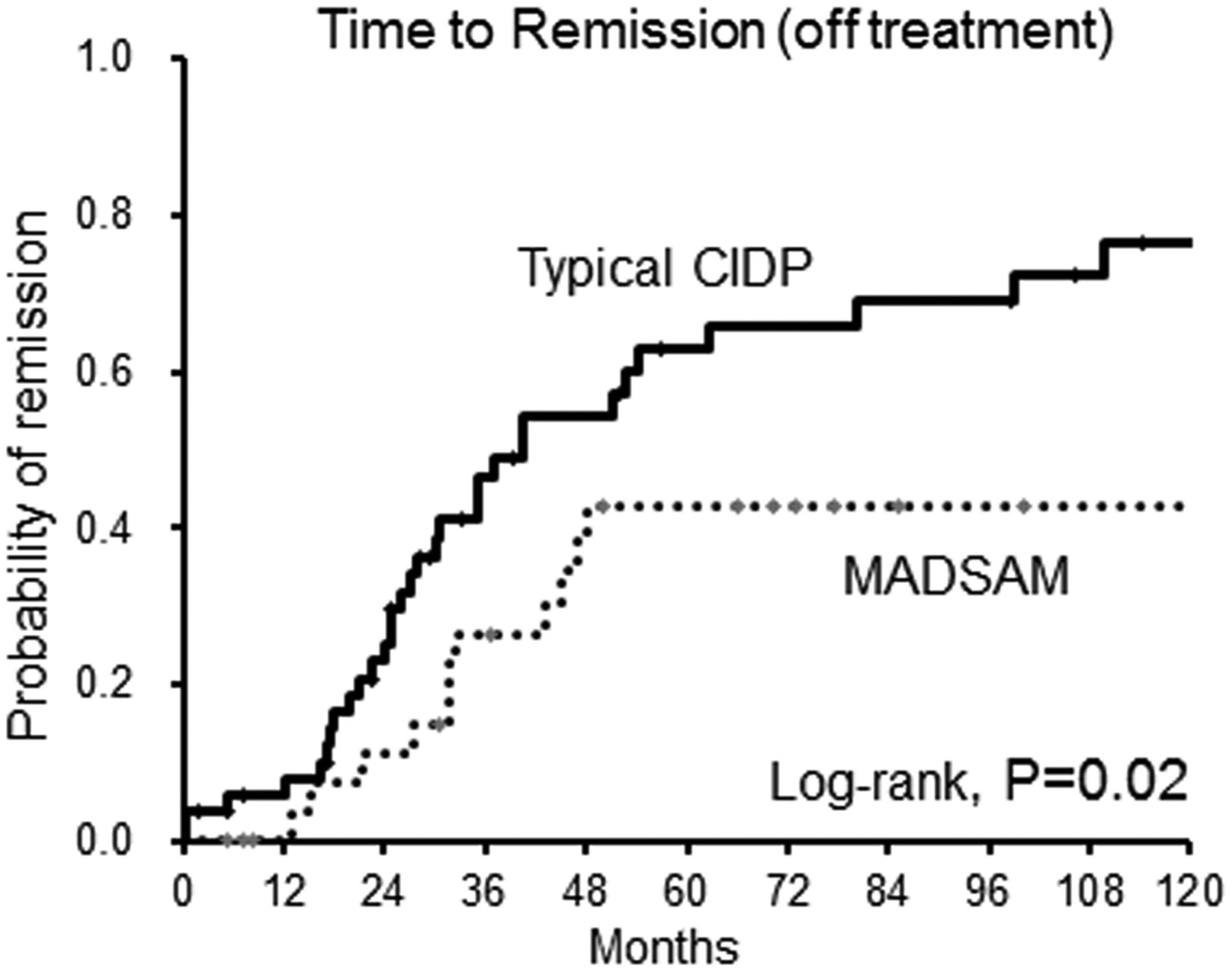
At the end of follow-up, 55% of the typical CIDP were in remission (Hughes grade 0–1), and 24% had partial remission (Hughes grade 2), whereas in patients with MADSAM remission was achieved in only 33%, and 40% had severe disability. In typical CIDP and MADSAM, muscle trophy was associated with poor response to treatment. Owing to the large variability of the follow-up period among the patients, we used the Kaplan-Meier analysis, and figure 3 shows the Kaplan-Meier curves for the probability of reaching clinical remission (Hughes grade 0 or 1, and off treatment). The long-term outcome was better for typical CIDP than for MADSAM. At 5 years after the start of treatment, 64% of the patients with typical CIDP reached clinical remission (Hughes grade 0 or 1), and 41% of the patients with MADSAM were in remission (p=0.02).
Kaplan-Meier curves to show time to remission (Hughes grade 0–1, and off treatment) in typical chronic inflammatory demyelinating polyneuropathy (CIDP) and multifocal acquired demyelinating sensory and motor neuropathy (MADSAM).
Azathioprine or cyclophosphamide were administered to only patients with MADSAM (n=7) who were refractory to the standard treatments, and did not appear to be clearly effective in all of them.
Profiles of patients with DADS
In this series, five patients (5%) had DADS. Owing to the small number of patients, a comparison of DADS and typical CIDP or MADSAM was not performed. Profiles of patients with DADS were similar to those with demyelinating neuropathy with anti-MAG antibodies (n=8), characterised by high age onset (median, 68; range, 60–77 years), male dominancy (5:0), chronic progression (median time to the first visit, 52 months), and refractoriness to corticosteroids and IVIg (none of them responded).
Discussion
Our results show that in the CIDP spectrum defined by EFNS/PNS criteria, typical CIDP and MADSAM neuropathy are the major clinical phenotypes and different electrophysiological features, treatment response and outcome between the two subtypes. This is the first study to directly compare the two subtypes, and total data suggest that the pathophysiology and immunopathogenesis are distinct between typical CIDP and MADSAM neuropathy.
Several studies have reported the frequency of CIDP subtypes in consecutive patients who fulfilled EFNS/PNS diagnostic criteria. A recent report from France has shown that among 146 patients with CIDP, 51% had typical CIDP, and 15% had MADSAM (Lewis-Sumner syndrome).8 In a report from the UK, 46 patients with CIDP had sensorimotor symmetric (presumably typical CIDP; 80.4%), MADSAM (15.2%) or DADS (6.5%).19 Compared with previous series, our CIDP cohort included smaller proportions of pure motor or sensory CIDP. As shown in figure 1, all patients with pure motor chronic demyelinating neuropathy had multifocal motor neuropathy according to published criteria.20 The definition of pure sensory CIDP has not been established; most of the reported patients with clinically sensory predominant or pure sensory show definite demyelinating motor nerve conduction abnormalities.8 ,21 Moreover, even if neuropathy is pure sensory at the first examination, motor weakness develops during the course of the disease;22 DADS neuropathy is a rare subtype constituting 5% of our whole patients with CIDP.
Our findings show that the two major CIDP phenotypes were typical CIDP (60%) and MADSAM (34%), and that the distribution patterns of nerve demyelination were substantially different among the two phenotypes. As shown in table 2, typical CIDP was characterised by a prolonged distal latency and low terminal latency index, suggesting demyelination predominance in the distal nerve terminals. In the distal nerve terminal and nerve roots, the blood–nerve barrier is anatomically deficient,23 ,24 although proximal lesions were not directly shown in this study. Previous studies have shown that the distal nerve terminals and roots are preferentially involved in Guillain-Barré syndrome,25–27 and our results support that this is the case for typical CIDP. Selective involvement in the distal nerve segments was found for 20% of our patients with typical CIDP, and involvement of the distal and intermediate nerve trunks was observed in 43%. We speculate that immune-mediated demyelination initially affects the distal nerve terminals/roots, and gradually extends to the nerve trunks with breakdown of the blood–nerve barrier by upregulated proinflammatory cytokines (eg, tumour necrotising factor-α) and complements.28 Future studies will be required to elucidate the pattern of progression in typical CIDP.
In the present study, sensory nerve conduction study results also indicate demyelination predominant in the distal nerve terminals of typical CIDP patients. We focused on the patterns of the abnormal median-normal sural sensory nerve responses, and this was seen in 53% of patients with typical CIDP, but only in 11% of those with MADSAM. Median sensory nerve conduction studies assess the most distal part of the nerve, whereas the intermediate portion was examined in sural nerve studies. It is established that distal demyelination should involve median sensory response more severely, and that sural response would remain normal. Similarly, ulnar sensory responses were also frequently abnormal, whereas superficial radial sensory responses were usually normal in our patients with typical CIDP (data not shown). Such a pattern of sensory nerve involvement is specifically seen in patients with CIDP or the Guillain-Barré syndrome.17 ,29 Combined with the favourable response to IVIg and plasmapheresis, antibody-mediated mechanisms would play a major role in the pathogenesis of typical CIDP.
In contrast, multiple sclerosis-like cellular immunity with breakdown of the barrier may be important in MADSAM neuropathy. Our results confirmed that MADSAM is characterised by a multifocal conduction block in the intermediate nerve trunk (seen in 62% of patients), and very rarely shows evidence of distal demyelination. The barrier may be disrupted by local activation of cell-adhesion molecules, inflammatory cytokines, matrix metalloproteinases or other inflammatory substances.30 The distribution of lesions in MADSAM is similar to that in multifocal motor neuropathy, but the pathophysiology is presumably different in the two disorders; in addition to the selective involvement of motor axons and the presence of serum anti-GM1 antibodies, the possibility of the altered membrane potentials and depolarisation block has been raised in multifocal motor neuropathy.31 Moreover, the treatment response is distinct; patients with multifocal motor neuropathy invariably respond to IVIg, but in this study only 37% of patients with MADSAM had improvement after IVIg. Conversely, corticosteroids are not effective for multifocal motor neuropathy, but this study showed that 72% of patients with MADSAM were responsive.
The treatment response and course of patients with MADSAM varied considerably among previous reports. In an original report by Lewis et al,32 two of the five patients improved after corticosteroids, and the remaining three had a chronic stable course and were therefore untreated. Later reports showed response to IVIg in 48–55% of patients, and to corticosteroids in 33%, whereas some patients worsened after steroid treatment.33 ,34 In general, 20–27% of patients with MADSAM had no response to standard treatments for typical CIDP, and experienced a chronic progression of the disease. Our findings showed that on the Kaplan-Meier analyses, only 41% of MADSAM had remission 5 years after the initiation of treatment. Novel treatment strategy is required for such patients with refractory MADSAM. Assuming that the possible pathophysiology is similar to that of multiple sclerosis, we suggest that agents targeting cell-mediated immunity, such as fingolimod and natalizumab, may be treatment options.
This study was retrospective and uncontrolled, and because of these limitations we must be cautious about the assessment of treatment response and pathophysiological hypotheses from the findings. Other limitations include the use of the Hughes functional grading scale that reflects lower limb function. However, this study included the largest number of patients with MADSAM, and direct comparison with typical CIDP revealed that the pathogenesis of the two CIDP subtypes appears to be distinct. Among the whole CIDP spectrum, the frequency of MADSAM may be higher than was previously thought, and a considerable number of patients with MADSAM are refractory to standard treatments for typical CIDP and require new therapies.
References
Supplementary materials
Supplementary Data
This web only file has been produced by the BMJ Publishing Group from an electronic file supplied by the author(s) and has not been edited for content.
Files in this Data Supplement:
- Data supplement 1 - Online supplement
Footnotes
Contributors SK, SI and SoM designed the study. MM, SaM, SS, MB and YS performed the examinations. SK and SoM drafted the manuscript. SK supervised the study.
Funding This study was supported in part by a research grant from the Ministry of Education, Culture, Sports, Science, and Technology of Japan (SoM and SK) and grants-in-aid from the Research Committee of Neuroimmunological Diseases, the Ministry of Health, Labour and Welfare of Japan (SK).
Competing interests None.
Patient consent Obtained.
Ethics approval The ethics Committee, Chiba University School of Medicine.
Provenance and peer review Not commissioned; externally peer reviewed.