Article Text

Statistics from Altmetric.com
Neurofibromatosis type 1 (NF1) is one of the commonest autosomal dominant disorders in man, affecting 1 in 3500 people. Consensus clinical criteria were defined in 19871 and revised and updated in 1997.2 Café au lait spots, axillary freckling, dermal neurofibromas, and Lisch nodules of the iris are the most common manifestations of this disorder. Most of the clinical symptoms of the disease are age dependent and considerable phenotypic variability has been described both between and within families.3,4 This genetic disorder is caused by mutations in the NF1 gene, one of the largest human genes, composed of 60 exons and spanning more than 300 kb of genomic DNA.5 The determination of the NF1 mutational spectrum has been complex owing to the large number of coding exons and the considerable mutational heterogeneity. Until recently, most diagnostic laboratories just offered linkage analysis for NF1 patients, which excluded diagnosis of the 50% of de novo cases. The use of techniques based on the analysis of NF1 mRNA greatly facilitated the number of mutations identified and NF1 screening efficiency, depicting a mutational NF1 spectrum.6–8 These studies highlighted the importance of splicing defects in molecular NF1 pathology and, despite most patients bearing unique mutations, they suggested the recurrence of several mutations.
Here we present our experience with the direct analysis of the whole NF1 coding region in 474 unrelated subjects suspected of having NF1. Mutations have been identified in 189 patients, 85 of them bearing recurrent mutations.
MATERIALS AND METHODS
Patients and families
Four hundred and seventy-four unrelated subjects suspected of having NF1 were analysed for mutations in the NF1 gene. Included in these 474 cases are 80 NF1 patients studied previously.6 Clinical data confirming NF1 diagnostic criteria were available in 201 (42%) of the subjects studied and in the remaining cases either no clinical data were provided or patients fulfilled only one diagnostic criterion. Patients with large deletions in the NF1 gene, previously detected by loss of heterozygosity (LOH),9 were excluded from this study. When available, blood samples of other family members were also obtained. All the participants were informed about the study and consent was obtained from all patients and their relatives.
DNA/RNA extraction
DNA was extracted from peripheral blood by the “salting out” method.10 Total RNA was extracted from peripheral blood lymphocytes using the Tripure isolation reagent (Boehringer Mannheim), according to the manufacturer's instructions.
Mutation analysis of the NF1 coding region by cDNA-SSCP/HD
Two to five μg of RNA were reverse transcribed using Superscript II reverse transcriptase (Invitrogen) and the entire NF1 cDNA was amplified in 10 overlapping fragments ranging in size from 636 to 1262 bp. These RT-PCR products were run in commercial 10% polyacrylamide gels (CleanGel DNA Analysis kit; Pharmacia Biosciences) and silver stained, in order to detect abnormal SSCP/HD patterns as previously described.6 The abnormal products were characterised by using an automatic sequencer (ABI PRISM™ 377). Finally, all the alterations were characterised at the genomic level by amplification and direct sequencing of the specific exon bearing the sequence change identified in the cDNA.
Statistical analysis
In order to determine whether mutations are equally distributed or not along the NF1 gene, we used the χ2 test to compare the observed frequency with respect to the expected one in every exon. Since mutations can be considered rare events, we assumed a Poisson distribution. We used SPSS software version 10.0 (SPSS Corp, Chicago, IL) and the test was evaluated using a significance level of 0.05.
Key points
-
The neurofibromatosis type 1 gene (NF1) has been described as bearing one of the highest mutation rates in the human genome. Half of the patients affected by NF1 are sporadic cases of the disease. Up to now it has been thought that most of these patients have private mutations which, in addition to the large size of the gene, has greatly hampered the definition of the mutational spectrum in NF1 patients.
-
We present here our experience of four years using the cDNA-SSCP/HD approach for mutational screening of the whole NF1 coding region. We have searched for NF1 mutations in 474 unrelated subjects suspected of having NF1. We have identified 142 different NF1 mutations in 189 patients. One hundred and four of these mutations have been found only once in this study, while the remaining 38 mutations have either been detected more than once in this study or have previously been published. Thus, we observed that 85 of 189 patients for which we identified a NF1 mutation (45%) harbour a recurrent mutation.
-
All detected alterations were characterised both at the genomic and RNA level. Considering the mutation effect in RNA processing, we have observed that 50% of patients harbour mutations that would lead to a recurrent alteration in mRNA. The detection of this high number of recurrent mutations could modify the routine genetic testing of the NF1 gene by performing a search for these recurrent mutations as a first analytical step.
RESULTS
Our centre has been offering mutational screening for the NF1 gene since 1999 once the cDNA-SSCP/HD approach was optimised for the analysis of the whole NF1 coding region in our laboratory.6 By using this technology, we have studied 474 unrelated subjects suspected of having NF1. We have identified 189 independent NF1 mutations in this sample comprising 142 different mutations (table 1). Eighty-five of these 189 unrelated patients (85/189, 45%) harbour 38 recurrent mutations (table 2) and the remaining 104 patients bear unique mutations (104/189, 55%). In all cases we have characterised both the DNA mutation and its effect at RNA level.
One hundred and forty-two different NF1 mutations and their effect on mRNA in 189 independent NF1 patients
Thirty-eight NF1 recurrent mutations in 85 independent NF1 patients
Description of mutations at the DNA level
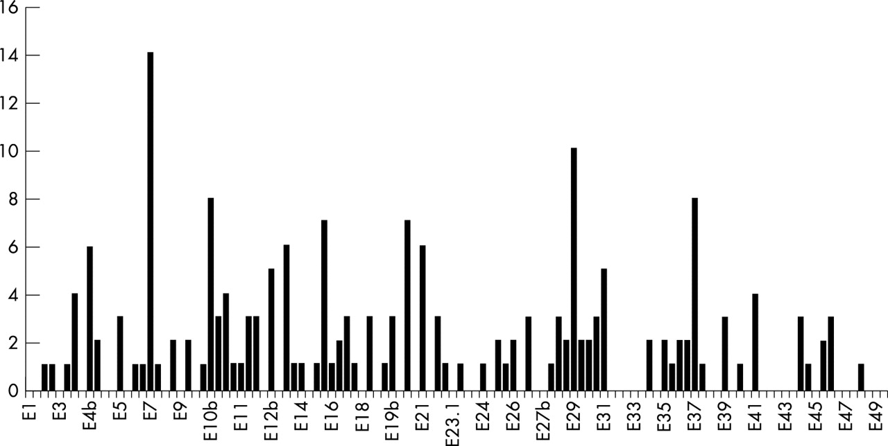
NF1 mutations identified in the present study are distributed along the NF1 gene (fig 1). However, there are eight exons/flanking introns in which mutations are represented more often (4b, 7, 10b, 13, 15, 20, 29, and 37), where 77 of the 189 mutations are located (41%), although they represent only 16% of the coding region. In order to test whether the mutations were equally distributed or not along the gene and assuming mutations as rare events, we have performed a χ2 test. This test showed that there are areas of the NF1 gene that have a greater tendency to accumulate mutations ( p<0.001).

Distribution of NF1 mutations in 189 neurofibromatosis type 1 patients. Exons and introns are represented consecutively on the X axis and the number of mutations identified on the Y axis.
Regarding the classification of the 142 different DNA mutations, we observed that half of them are frameshift mutations, 31% affect the splicing consensus sequences, 9% are nonsense mutations, 8% are missense mutations, and only 2% represent amino acid deletions. Twenty-seven percent of these mutations were recurrent (38/142).
Mutation effect on mRNA processing and the protein
When we studied the effect of these 142 mutations on mRNA processing, we obtained a different classification for the mutations. The percentage of mutations altering the correct splicing of the NF1 pre-mRNA increased to 40% since, apart from the mutations affecting the consensus splicing sequences, we also found three frameshift, four nonsense, and five missense mutations that produced an aberrant NF1 pre-mRNA splicing. If we consider this effect, 38% (54/142) of the identified mutations would lead to a recurrent effect on mRNA and are present in 50% of the patients. In relation to the putative effect of mutations at the protein level, only six mutations (4%) are not predicted to modify the size of neurofibromin. The remaining mutations would affect the neurofibromin size, 19% would produce an in frame protein with a slightly larger or smaller size than the authentic neurofibromin, and 77% of the mutations would lead to a truncated neurofibromin.
Recurrent mutations
Eighty-five of the 189 independent NF1 patients in which the germline mutation has been identified (45%) harbour 38 different recurrent mutations (table 2). These mutations have been found in different unrelated patients of our set and/or have been previously published by other groups. In our population the most frequent mutations are two nonsense and two missense mutations, although interestingly all of them alter the correct mRNA splicing. The commonest mutation identified is 910C>T (R304X), which was found in seven independent patients. This mutation causes skipping of exon 7 and has been reported in several NF1 mutational studies.7,8,11 This mutation represents 8% of the recurrent mutations and 4% of the total of mutations identified. The commonly reported 6792C>A (Y2264X) mutation, which produces exon 37 skipping, has been identified in six patients, being the second most frequent recurrent mutation in our population. Two mutations were found in five unrelated patients (1885G>A and 5546G>A). Mutation 1885G>A inactivates the 3` consensus splice site and a cryptic 3` splice site is used instead, producing the deletion of 41 nucleotides of exon 12b. Mutation 5546G>A causes the inactivation of the 5` splice site which leads to the skipping of exon 29.
Identification of amino acid variants in the coding region
By using the cDNA-SSCP/HD assay we have detected four nucleotide substitutions that produce a change of amino acid in the affected position but which are not associated with the disease (table 1). Two changes are located in exon 29, R1825W (5473C>T) and R1809C (5425C>T), one in exon 4b, D176E (528T>A), and one in exon 21, N1229S (3686A>G). These changes were found in four sporadic NF1 patients and were also present in other unaffected relatives in their families. In the patient carrying the N1229S change, we characterised the causative NF1 mutation (1466A>G, exon 10b); however, in the other three patients we were unable to detect any other nucleotide alteration. A priori these changes should be considered as rare variants owing to the fact that we have only identified them once in the sample of 474 independent patients analysed in this study. Variant D176E has previously been reported four times as a polymorphism in the German population,7,12 while the other variants are described for the first time in this report.
Phenotype-genotype correlation
No clear correlation has been found between a specific NF1 mutation and a particular clinical feature. First of all, it has to be taken into account that in a large number of cases we have little clinical information owing to the fact that samples are referred to our laboratory from all over the country and from different clinical services. It is interesting to note that 66 of the studied patients were children with healthy parents aged from several months to less than 10 years. Most of these children have only café au lait spots and in some cases freckling was also reported. Although some of them do not fulfil the NF1 NIH consensus criteria, we have detected a NF1 mutation in 20 of them (30%), confirming in these cases the affected status of the child. Considering the patients from whom clinical data were available, several findings were observed.
Café au lait spots and dermal neurofibromas
In general terms, although most patients present these age dependent traits, the quantitative information has been very poor in the vast majority of them. However, as we have previously described,6 we have found the same mutation in patients of similar age with a very different number of lesions. This has been mainly seen in the cases with the most prevalent mutations (910C>T, 6792C>A, 1885G>A, and 5546G>A) in which it has been possible to compare a larger number of patients.
Plexiform neurofibromas
We have detected the NF1 causing mutation in 15 of 25 unrelated patients in which the presence of plexiform neurofibromas was reported. The majority of these mutations will produce a truncated protein (13/15) and just two would produce a shorter in frame neurofibromin (IVS10b+1G>A and IVS18+5G>C). Considering these 15 patients, mutations 1466A>G and R681X are both present in two of them and two other patients have different mutations in exon 20 (3403-3404delTC and 3456-3459delACTC). The remaining patients harbour different recurrent (1541-1542delAG, 3822-3823delTC, R1947X, IVS45+790C>G) or unique (IVS10b+1G>A, 1756-1759delACTA, IVS17+2insT, IVS18+5G>C, IVS22+1G>C) mutations. Most of these mutations are outside the GRD although two are within it.
Scoliosis
The NF1 mutation was characterised in nine of the 18 patients with scoliosis. Interestingly, four of these patients have the same mutation in exon 10b (1466A>G), another has a mutation in intron 10b (IVS10b+1G>A), and the other four harbour different mutations (IVS7+1G>A, IVS30+322A>G, IVS37+2T>G, and IVS45+790C>G). None of these mutations is in the GRD.
Optic glioma
Among 20 patients with optic glioma, we have detected the NF1 mutation in 12. Most of these mutations are in the first exons of the NF1 gene and three of them are located in exon 7 (1019insT, 989insC, and 998insA); all mutations are different and will produce truncated proteins. Apart from mutations in exon 7, the other mutations found were: 590delC, 1466A>G, IVS10a-9T>A, 1541-1542delAG, 1756-1759delACTA, 2173insT, 5351insC, 7204-7205delCA, and 7702C>T.
Learning disabilities and mental retardation
Mental retardation has only been described in two patients harbouring mutations R681X and IVS30+322A>G. Among 16 patients described with learning disabilities we identified the NF1 mutation in six. All of them carry different mutations that produce truncated proteins (989insC, IVS10c-2A>G, IVS15-16A>G, IVS23.1-2A>G, and 5303delA), except patient 99-135 who harbours a missense mutation in the GRD domain (4493G>A, G1498E).
Detection of abnormal transcripts not caused by NF1 mutations
The routine analysis of the NF1 cDNA by SSCP/HD leads to the identification of several aberrant transcripts expressed in blood at a low proportion in comparison to the wild type transcript. When the genomic sequences corresponding to these transcripts were obtained, no DNA change was identified. The sequence of these abnormal transcripts suggests that they correspond to several previously described aberrant splice forms, such as the insertion of a cryptic exon (exon 4a-2),13,14 the skipping of some constitutional exons such as exon 2015 or exon 43,15–17 and also the presence of one of the alternative NF1 isoforms consisting of the insertion of exon 23a.18 Most of the samples showing these abnormal transcripts were samples sent to our laboratory from a long distance away and therefore the RNA was extracted several days after drawing of the blood.
DISCUSSION
Mutational NF1 screening for diagnostic purposes has been reported to be a difficult task owing to the size of the gene and to the absence of clear mutational hot spots. In the present study, we have observed that recurrent mutations are more common than previously described, being present in 45% of the patients in whom the NF1 causative mutation has been identified. This percentage rises to 50% if we consider the effect of mutations on mRNA processing. This result has important implications for the future design of NF1 genetic tests and introduces the possibility of developing rapid tests focused on searching for these recurrent mutations as a first step in the routine diagnostic procedure. However, the recurrence rate calculated from the population studied here should be considered with caution since this population includes a large number of patients for whom either no clinical data were available or who did not fulfil the NF1 diagnostic criteria. Therefore, an accurate final estimation of the mutation detection rate is not possible and, hence, a detection bias for certain mutations cannot be excluded.
Here we report the NF1 mutational analysis of 474 unrelated subjects suspected of having NF1 in whom we have detected 189 mutations. The mutations are scattered along the gene with some exons having a higher number of mutations, corresponding to exons harbouring recurrent mutations. We have found that 77 of the 189 mutations (41%) are located in just eight exons/flanking introns which represent only 16% of the coding region. The chi-square test gives statistical significance for a non-Poisson distribution of mutations, proving that they are over-represented in these exons (p<0.001).
Mutation 910C>T (R304X) in exon 7 is the commonest mutation detected in our population and this exon/flanking intron is the most mutated in our sample, in 15 out of 189 patients (8%). In addition both exons/flanking introns 10b and 29 are mutated in 11 patients each (6%). Our results agree with published reports on the absence of mutations in the alternative exons 9br, 23a, and 48a, but failed to find mutations in exons 23.2 and 27a which have previously been described as exons harbouring recurrent mutations.7,8
The predicted effect of the mutations on neurofibromin size is variable, although a great proportion of them will lead to the formation of a truncated protein. However, it is interesting to note that almost half of the mutations affecting RNA splicing would induce the formation of in frame transcripts resulting in a putative protein slightly shorter or longer than the authentic neurofibromin, but not truncated. We have identified six missense mutations and three deletions of amino acids (6%); this proportion is in agreement with data presented previously.7
Moreover, four polymorphic variants have been identified (D176E, N1229S, R1809C, and R1825W). These changes were found in sporadic patients and did not segregate with the disease since they are present in several unaffected relatives of the families. Variant N1229S is located in the GAP related domain and it is found in a patient in whom a NF1 causative mutation has been identified (recurrent mutation 1466A>G). This patient is a 27 year old woman with the typical clinical features of NF1 including café au lait spots, neurofibromas, Lisch nodules, and optic glioma. On the other hand, we were unable to detect any other change in the other three patients. The patient with variant D176E is a 22 year old woman with a classical NF1 presentation; this variant has previously been described as a polymorphism in four NF1 German patients, thus it should also be considered a polymorphism in this patient.7,12 The remaining two patients are children younger than 5 years with a clinical suspicion of NF1 but without a firm diagnosis.
The lack of clinical information for most of the patients referred to our laboratory makes it impossible to provide final data about efficiency in NF1 mutation detection. Although at first glance the mutation detection rate is lower than in our previous work,6 it should be taken into account that in the pilot study only patients fulfilling NF1 consensus diagnostic criteria were included. Since our centre offers direct genetic analysis of the whole NF1 coding region all over Spain, not only classical NF1 cases are referred but also clinically ambiguous patients, usually showing only one criterion of the disease. For instance, a considerable number of patients studied here (66 out 474, 14%) are children aged from a few months to less than 10 years, most of them with only café au lait skin spots. These samples are mainly sent to our laboratory from paediatric services in order to confirm the probable diagnosis of NF1. In 20% of these patients we have detected the NF1 mutation. For mutational screening by the cDNA-SSCP/HD approach we discarded patients bearing large NF1 deletions detected by LOH or by FISH analysis, which represent around 5% of the NF1 mutations in our population.9 The approach used here has several limitations. Some single nucleotide mutations may be missed in the SSCP/HD analysis owing to the large size of the amplified fragments. Another important mechanism hampering mutation detection when using a RNA based approach is nonsense mediated mRNA decay (NMD), which could cause an under-representation of the mutated transcript.8,19 Also, with this methodology, deletions involving several exons would be lost. Mechanisms reported in other genetic disorders such as large duplications or inversions cannot be ruled out as responsible for the remaining unidentified NF1 mutations. Moreover, mutations located in the 5` or 3` untranslated regions should also be considered. Lastly, somatic mosaicism could be underlying some of the sporadic cases in which a mutation has not been identified in blood samples.
As previously described, special care has to be taken when an RNA approach is used for diagnostic purposes. We have detected a large number of aberrant transcripts expressed in a low proportion compared to the normal one that could be misinterpreted as a result of the pathogenic mutation. However, most of them have been shown to be illegitimate transcripts produced by cold shock stress. This phenomenon is exacerbated when RNA is not extracted immediately after blood collection.13–16 Unfortunately, 90% of samples are sent from other hospitals making it impossible to know how much time has elapsed after venepuncture. The establishment of short term cell cultures before RNA extraction and the addition of inhibiting substances to reduce NMD could be very useful in solving this problem and would improve the efficiency of the analysis.8 With these possible sources of diagnostic error, to avoid mistakes in the final diagnosis, alterations detected in the cDNA must always be confirmed at the genomic level. This characterisation at both levels also provides interesting information, as we describe here, that some missense, nonsense, and frameshift mutations produce splicing alterations instead of the effect predicted for the mutation at the DNA level. These results have also to be taken into account in the design of functional studies using mutated protein models.
As has been described in published reports, it is very difficult clearly to identify any relationship between a mutation and a determined feature of the disease. In the work presented here, the establishment of any relation has been very limited because in a large number of cases we have not been able to obtain good clinical data on the patient(s). Moreover, the fact that café au lait spots and neurofibromas are age dependent quantitative traits has greatly hampered the possibility of making comparisons. However, several trends reported previously by our group6 seem to be confirmed, such as the large number of mutations in exon 10b in patients with scoliosis and the fact that all patients with optic glioma harbour mutations that would produce a putative truncated neurofibromin. In contrast, it has been impossible to shed light on the putative relation of harbouring mutations producing altered in frame transcripts and a low number of neurofibromas. Nevertheless, the most probable explanation for the extreme clinical heterogeneity in NF1 is the role of modifier genes determining the expression of the clinical manifestations.3,4 Another possibility could be that mutations altering the correct splicing of the NF1 pre-mRNA could lead to variable levels of the abnormal transcript in different patients bearing the same mutation.6 In order to elucidate the contribution of this event in NF1 aetiology, we are currently performing a study based on semi-quantitative PCR. Although our data are very preliminary, we have observed differences between patients in the percentage of mutated NF1 transcripts versus wild type (E Pros, unpublished data).
In conclusion, the routine analysis of the NF1 gene for diagnostic purposes shows that recurrent mutations are more common than previously expected, accounting for more than 40% of identified mutations. The new technological advances such as the microarray technology could be very useful for the genetic screening of the NF1 gene; a chip harbouring the recurrent mutations would facilitate the molecular diagnosis of the disease. However, new approaches should be developed in order to identify mutations in the remaining patients. A combined approach such as the one described by Messiaen et al8 based on a cascade of complementary techniques could be a good methodology although it is very laborious and expensive for routine analysis of hundreds of samples per year. Lastly, a method designed for the detection of multi-exon deletions should be considered in order to evaluate the role of this type of mutation in the molecular pathology of NF1.
Acknowledgments
The first three authors contributed equally to this work. The authors wish to thank all the patients and family members who participated in this study and all the clinicians for sending the family samples. We are very grateful to Rafael de Cid for the statistical calculation. This work was supported by grants of the Fondo de Investigaciones Sanitarias de la Seguridad Social (01/1475), the Ministerio de Ciencia y Tecnología (SAF2002-00573), and the Institut Català de la Salut.