Article Text

Abstract
Two patients with moderate Huntington’s disease (HD) received bilateral fetal striatal allografts. One patient demonstrated, for the first time, increased striatal D2 receptor binding, evident with 11C-raclopride positron emission tomography, and prolonged clinical improvement over 5 years, suggesting long term survival and efficacy of the graft. The other patient did not improve clinically or radiologically. Our results indicate that striatal transplantation in HD may be beneficial but further studies are needed to confirm this.
Statistics from Altmetric.com
Huntington’s disease (HD) is an incurable neurodegenerative disorder characterised by progressive neuronal death, particularly in the striatum. One strategy to treat this relentless progression is to replace the lost or dysfunctional striatal neurons through fetal striatal allografts. This approach was successful in animal striatal lesion models of HD,1 2 heralding several human pilot trials. A French study showed clinical and 18F-fluorodeoxyglucose positron emission tomography (PET) stabilisation or improvement for up to 4 years in three out of five patients with HD transplanted with fetal striatal allografts.3 However, 18F-fluorodeoxyglucose PET reflects cellular metabolism and synaptic activity, which may be increased by a glial reaction to grafting and does not specifically indicate graft survival. Another study4 found no clinical or PET improvement in seven transplanted patients despite postmortem evidence of graft survival with striatal tissue in one patient.5 In the latter study, the patients were more advanced and complications from the neurosurgical procedure were relatively common. The researchers also adopted a different tissue preparation method. Neither study demonstrated specifically in vivo survival of striatal tissue in a patient who had clinically improved.
In this study, we report on two patients with HD transplanted with human fetal allografts and followed up for over 5 years. In one of them, there was prolonged clinical improvement and increased striatal D2 receptor binding, evident with 11C-raclopride (RAC) PET, suggesting long term survival and efficacy of the graft.
SUBJECTS AND METHODS
The study received approval from the local research ethics committees. All subjects gave written informed consent in accordance with the Declaration of Helsinki.
Subjects
Two patients with HD received fetal striatal allografts. Subject No 1 (male, CAG repeat 43) was diagnosed with HD at the age of 46 years when he developed chorea, aggressive behaviour and depression. He was treated with reboxetine 8 mg/day which was maintained throughout the study period. Subject No 2 (female, CAG repeat 43) presented at the age of 49 years with chorea and gait instability. She was not on any medication prior to the transplantation. Disease duration at the time of transplantation was 6 years for subject No 1 and 4 years for subject No 2. Tables 1 and 2 show their baseline characteristics.
Tissue preparation and neurosurgical procedures
Informed consent to collect fetal tissue of 9–10 weeks’ gestational age was obtained from maternal donors who had already decided to terminate their pregnancy. The whole ganglionic eminence, dissected from striatal tissue of two to three fetuses, was implanted as a suspension into the target striatum on the same day as collection. The implantation was made along four trajectories into each target striatum—one in the caudate nucleus, with further tracks in the anterior, middle and posterior putamen. The contralateral striatum was implanted within 2 months. Preoperative MRIs were done to guide stereotactic implantation. Patients were immunosuppressed from the day of grafting with prednisolone for 1 month and ciclosporin for 1 year (see appendix online for further details).
PET scanning
RAC PET was used to image striatal D2 receptor binding, as recommended by CAPIT-HD.6 Increase in striatal RAC binding following striatal transplantation indicates graft survival and correlates with functional improvement in animal models of HD.7 PET scanning and measurement of striatal RAC binding potential were performed as previously described8 (see appendix online). Neither of the patients was receiving neuroleptic medication.
Clinical and neuropsychological assessments
The subjects were assessed clinically using the Unified Huntington’s Disease Rating Scale (UHDRS) and a battery of neuropsychological tests sensitive to HD pathology (table 1). All clinical assessments were performed, unblinded, by IR up to the 36 month time point, and thereafter by RAB because of a change in personnel and follow-up arrangements. Both are experienced movement disorders specialists.
Follow-up
PET scanning and clinical assessments were performed at baseline and at regular intervals following surgery—initially 6 monthly, and thereafter every 12–18 months.
HD controls
A group of six patients with HD (five men, mean age 48.6 (SD 7.1) years, disease duration 3.0 (1.3) years, CAG repeat 44 (3.7)) who did not undergo surgery were used as controls for the two surgical subjects. They also underwent serial RAC PET and clinical assessments. No sham surgery was performed as it was deemed inappropriate by the ethics committee, particularly in the setting of a pilot study.
RESULTS
Subject No 1
Clinical course
The patient had no perioperative complications. However, he developed moderate renal impairment while receiving ciclosporin which consequently had to be discontinued after 3 months. Overall, there was a gradual but significant improvement in his condition, reported both subjectively and with clinical scales. His UHDRS motor score (decrease from 54 to 8 over 5 years), functional capacity, Beck Depression score and, to a lesser extent, cognitive functions (eg, verbal fluency) all showed marked improvement in the first 3 years after transplantation, and have remained stable thereafter (table 1).
PET findings
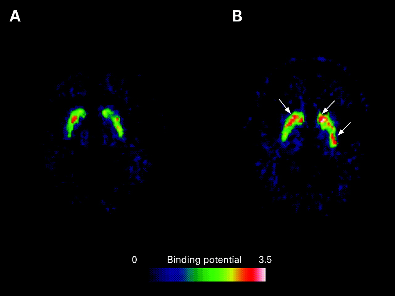
The striatal RAC binding potential of subject No 1 increased by 23% at 6 months post-transplantation but then declined at a rate similar to the HD controls (figs 1, 2). At 2.5 years post-transplantation, his mean (left and right averaged) striatal RAC binding remained higher than baseline.

Subject No 2
Clinical course
The patient had no perioperative or postoperative complications. Her clinical condition did not change markedly after transplantation. However, 2 weeks after completing her 12 month course of ciclosporin, she experienced increasing gait disturbance which resulted in a fall and head injury. She developed neck stiffness, pyrexia and a reduced level of consciousness. MRI at this time showed enhancement and oedema around the transplant tracks. CSF examination revealed a lymphocytosis with raised protein and decreased glucose levels, but no organisms were isolated. She was treated empirically with broad spectrum antibiotics and antituberculous therapy. She recovered and returned to her preoperative level of functioning before deteriorating at a rate that is similar to that seen in non-transplanted patients with HD.
PET findings
Mirroring the patient’s clinical outcome, her RAC binding did not improve post-transplantation and continued to decline at a rate similar to the HD controls.
Table 1 shows the full clinical scores and PET results. Some data points were missing during the changeover of personnel.
DISCUSSION
We have reported for the first time, in one of two subjects with HD undergoing fetal striatal allografting, prolonged and concomitant improvement in motor function and striatal D2 binding, implying survival and development of the graft. Subject No 1 is now 12 years into his disease and continues to be independent in all aspects of activities of daily living. It is possible that the resolution of his depression, as reflected by the improvement in his Beck Depression scores, may account for some of his clinical improvement. However, neither this nor placebo effects could explain the prolonged improvement in PET and the subsections of his UHDRS motor scores, including chorea (see appendix online) over 5 years. Furthermore, the profile of cognitive improvement points not to a global change, as would be expected in someone with resolving depression, but rather shows changes more in frontostriatal tasks.
Striatal RAC binding in subject No 1 increased by 23%, 6 months post-implantation, which correlated with clinical improvement, suggesting survival and function of the graft. The binding declined gradually thereafter, most likely indicating the progression of his HD and associated loss of intrinsic striatal projection neurons. His continual clinical improvement may have been sustained by further functional integration of the surviving graft, despite a declining overall number of striatal neurons. The apparent radiological–clinical dissociation at the later stage does not invalidate RAC PET as a radiological marker of graft survival and function, but merely reflects the additional effect of HD progression and its impact on the intrinsic striatal neurons.
In contrast, subject No 2 and the non-grafted HD controls continued to deteriorate. Subject No 2 had an acute meningoencephalitic illness which may have been related to a skull fracture sustained following a fall. The lack of clinical and PET improvement post-implantation implies that the graft failed to survive or differentiate appropriately. Suboptimal fetal tissue handling or the potential adverse effects of the meningoencephalitis on the graft are probable causes for the lack of improvement. An alternative explanation may be ongoing low grade chronic graft rejection which can become manifest after cessation of immunosuppressants, as recently reported in another HD transplantation study.9 We did not measure HLA antibodies in this patient to look for evidence of alloimmunisation.
Fetal striatal transplantation is a therapy that may alter the progression of striatally mediated aspects of HD, rather than a cure. Its effect on the cortical pathology of HD may be minimal, although improvement in movement related cortical activation has been seen in patients with Parkinson’s disease receiving striatal dopaminergic grafts.10
It is difficult to make a conclusive statement based on the small number of subjects studied here and the absence of blinded assessments and matched controls in a disease with high variability. Nevertheless, our results, together with the French series,3 show that fetal striatal transplantation may be beneficial in some patients with HD in the longer term. It is not possible to predict which patients might benefit from this procedure at present. A larger scale randomised controlled HD striatal transplantation study currently underway11 may provide a more definitive answer on the efficacy of this procedure in a disease which has no effective treatment.
REFERENCES
Footnotes
-
The appendix is published online only at http://jnnp.bmj.com/content/vol79/issue8
-
Funding: The study was funded by the Medical Research Council, UK. YFT was funded by the Wellcome Trust (GR071659AIA).
-
Competing interests: None.
-
Ethics approval: The study received approval from the local research ethics committees.