Article Text

Abstract
Background and purpose Infarct volume and age are strong predictors of outcome in patients with stroke. We aimed to determine the impact of infarct volume on outcome according to age.
Methods Consecutive patients with acute stroke with documented internal carotid artery/middle cerebral artery occlusion who underwent endovascular procedures were studied. Patients were categorized in three age groups: <70 years (G1), 70–79 years (G2), ≥80 years (G3). The Alberta Stroke Program Early CT score (ASPECTS) was graded on initial CT. Time of successful recanalization (Thrombolysis In Cerebral Infarct (TICI) ≥2b )and good outcome at 3 months (modified Rankin Scale score ≤2) were recorded. Infarct volume was measured on the 24 h control CT.
Results A total of 214 patients were studied (G1: 68; G2: 74; G3: 72). For all patients the mean infarct volume was 94.7±127 mL; 35.6% had a good outcome. We observed larger infarct volumes in patients with a bad outcome in each age group (G1: 22 vs 182 mL, p<0.01/G2: 22 vs 164 mL, p<0.01/G3: 7.6 vs 132 mL, p<0.01). However, the target cut-off infarct volume that better predicted a good outcome decreased as age increased: G1: 49 mL (sensitivity 80%, specificity 92.6%); G2: 32.5 mL (sensitivity 80%, specificity 81%); G3: 15.2 mL (sensitivity 81.3%, specificity 86.7%). Overall, after adjusting for age, occlusion location, baseline NIH Stroke Scale score and infarct volume, the only predictor of a good outcome was achieving a final infarct volume less than the age-adjusted target (OR 5.5, 95% CI 1.6 to 18.8; p<0.01). The probability of achieving an infarct volume less than the age-adjusted target decreased according to baseline ASPECTS, time and degree of recanalization.
Conclusions Age-adjusted infarct size might represent a powerful surrogate marker of stroke outcome and further refine the predictive accuracy of infarct volume on prognosis in patients with stroke undergoing endovascular treatment. This information may be used in the design of new trials to individualize selection criteria for different age groups.
- Angiography
- CT
- Intervention
- Stroke
- Thrombectomy
Statistics from Altmetric.com
Introduction
The rationale for recanalization therapies in acute ischemic stroke is to preserve the brain from ischemic damage and development of irreversible infarct lesion. The paradigm ‘time is brain’1 refers to the fact that shorter time of ischemia is generally related to smaller final infarct volumes. Recent studies have shown that successful recanalization leads to improved functional outcomes through a reduction in final infarct volumes.2–4 These studies identified patient age and final infarct volume as powerful independent predictors of outcome.
The benefits of endovascular procedures for acute stroke in older patients are under discussion,5 ,6 and more restrictive selection criteria should probably be applied as patient age increases.7 Age may determine functional recovery after a stroke since it is associated with a higher incidence of comorbidities and probably to a lower capacity of neurorestoration or plasticity.8 The impact of infarct volume on long-term stroke outcome may therefore be markedly affected by the age of the patient. In this study we aimed to determine the impact of infarct volume on outcome according to patient age.
Methods
Consecutive patients with acute stroke treated with endovascular procedures within 8 h from onset prospectively included in the database of a major comprehensive stroke center were divided into three groups according to age: <70 years (G1), 70–79 years (G2), ≥80 years (G3). Baseline characteristics, occlusion location based on initial angiogram and outcome measures were prospectively collected. Only patients with a baseline modified Rankin Scale (mRS)score <2 were included in this study.
Patient selection
All patients presenting with acute ischemic stroke symptoms underwent a baseline head CT scan and CT angiography (CTA). In general, as a first step, patients with anterior circulation stroke were selected for intra-arterial therapy if the CT scan showed an Alberta Stroke Program Early CT score (ASPECTS) of ≥7 and a major intracranial occlusion on CTA (terminal internal carotid artery (ICA) or middle cerebral artery (MCA) M1 or M2 segments). Most patients presenting >4.5 h after symptom onset (or unwitnessed onset) underwent additional imaging studies (CTA/CT perfusion) or MRI/MR angiography and were considered for interventional therapy based on assessment of the mismatch between the extent of infarcted brain relative to the extent of threatened but viable brain. The minimum amount of mismatch necessary for treatment selection varied according to patient-specific considerations and stroke neurologist/interventionalist-specific practice patterns. In general, patients with a core volume of less than one-third of MCA territory in the presence of ICA or M1 occlusion and corresponding clinical deficit (NIH Stroke Scale (NIHSS) score >8) or severe perfusion deficit (time to peak >6 s) involving two-thirds or more of the MCA territory were considered potential treatment candidates. Computer-generated volumetric analysis was not available and manual calculation of volumes is a too lengthy process to be useful for selection in the setting of acute stroke. Therefore, for treatment purposes, volumes were estimated based on visual mismatch. All patients were treated within 8 h of symptom onset.
Intra-arterial treatment protocol
The general approach regarding intra-arterial treatment at our center has been described previously.9 In most cases the procedure was performed under conscious sedation. Subjects with airway compromise or uncontrollable agitation were intubated before the procedure. According to device availability, patients were respectively and primarily treated either with intra-arterial tissue plasminogen activator (n=78) and/or a clot retriever device: Merci (n=14), Trevo (n=36) (Concentric Medical, Mountain View, California, USA), Solitaire (n=61; ev3 Endovascular, Plymouth, Minnesota, USA), pREset (n=3; Phenox GmbH, Germany). Manual aspiration was also used as a primary option (n=22) or as an adjunct to other approaches. The time and degree of recanalization were recorded; successful recanalization was defined as Thrombolysis In Cerebral Infarction (TICI) ≥2b.
Infarct volume calculation
A 24–36 h follow-up CT scan was performed; patients for whom this CT scan was not available were excluded from the analysis. Post-treatment infarct volumes were calculated by a vascular neurologist (EM) blinded to all other data. The acute infarct was determined by visual inspection on the follow-up CT scan. Final outlines were manually edited. If present, hemorrhagic changes were incorporated in the final infarct volumes. Symptomatic hemorrhage was defined as a CT-documented hemorrhage that was temporally related to deterioration in the patient's clinical condition in the judgment of the clinical investigator.10
Patient follow-up
On discharge, patients were scheduled for follow-up appointments in the stroke neurology clinic at 90 days. The mRS score was recorded at this time by an mRS certified neurologist. If patients were physically unable to present for follow-up, they or the next of kin were contacted by a vascular neurologist or other mRS certified investigator and the mRS score was obtained over the telephone. A good clinical outcome was defined as mRS score ≤2.
Statistical analysis
Descriptive and frequency statistical analysis was obtained and comparisons were made using the SPSS V.17.0 statistical package. Statistical significance for intergroup differences was assessed by the Pearson χ2 test or the Fisher exact test for categorical variables and the Student t test and analysis of variance for continuous variables. When indicated, Mann–Whitney U tests and Spearman tests were used. To calculate the sensitivity and specificity of infarct volume cut-off points to predict a good outcome, a receiver operator characteristic curve was configured. Multivariate logistic regression analyses were performed to determine factors that could be considered independent predictors of a good clinical outcome. Variables showing p<0.1 in univariate analysis were included in the multivariate model. A p value of <0.05 was considered significant for all tests.
Results
A total of 214 patients were studied (G1: 68, G2: 74, G3: 72). Older patients were more frequently women, had more hypertension or atrial fibrillation and presented with higher systolic blood pressure; other baseline characteristics were similar (table 1). For all patients the mean infarct volume was 94.7±127 mL; 35.6% had a good outcome. Mean infarct volumes were 96±131 mL for G1, 109±139 mL for G2 and 87±116 mL for G3 (p=0.71). Older patients tended to have a less good outcome (45.8% in G1, 33.5% in G2 and 27.3% in G3; p=0.1).
Patient characteristics according to age group
Infarct volumes
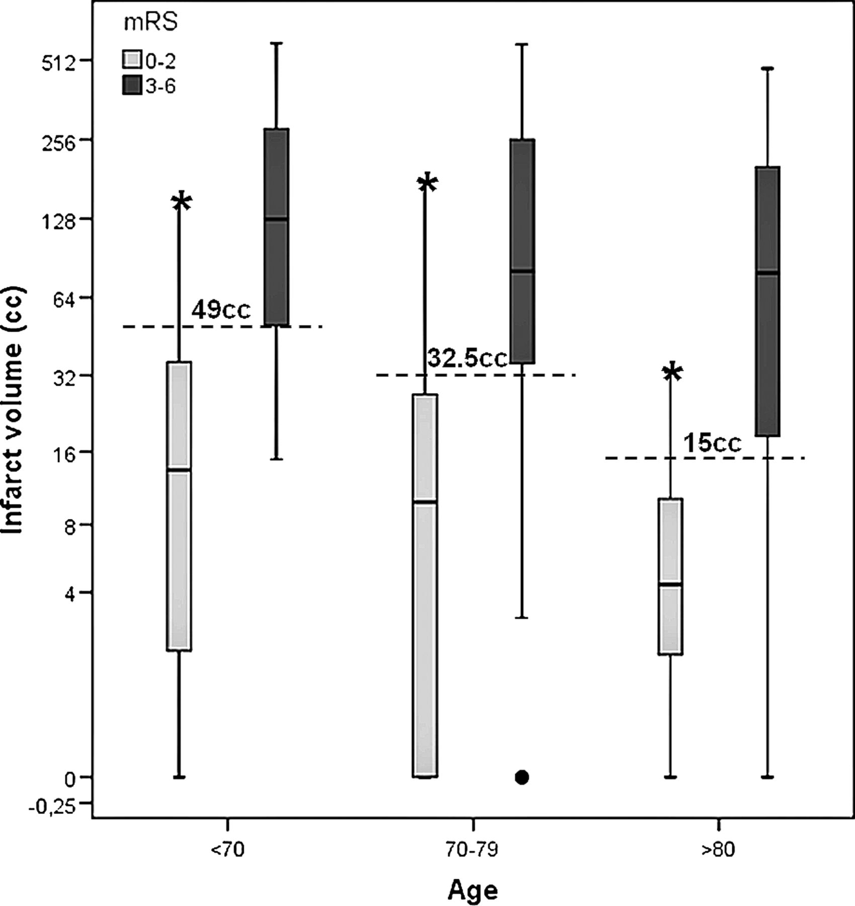
Patients with a poor outcome had larger infarct volumes overall (mRS 0–2: 21±35 mL vs mRS 3–6: 159±150 mL; p<0.01) and within each age group (G1: 22 vs 182 mL, p>0.01/G2: 22 vs 164 mL, p>0.01/G3: 7.6 vs 132 mL, p>0.01) For all patients the cut-off infarct volume that better predicted a good outcome was 29 mL (sensitivity 81.4%, specificity 79.4%). However, the cut-off infarct volume that better predicted a good outcome decreased as age increased (G1: 49 mL (sensitivity 80%, specificity 92.6%); G2: 32.5 mL (sensitivity 80%, specificity 81%); G3: 15.2 mL (sensitivity 81.3%, specificity 86.7%); figure 1).

Infarct volumes according to age group and 3-month outcome. Age-adjusted infarct volumes that better predicted good outcome. mRS, modified Rankin Scale.
On univariate analysis (table 2), in addition to infarct volume (p<0.01), variables associated with a good outcome were age (p=0.05), glycemia (p=0.05), use of intravenous tissue plasminogen activator prior to the procedure (p=0.01), terminal ICA occlusion (p<0.01), baseline NIHSS score (p<0.01), baseline ASPECTS score (p<0.01), time to initiate the procedure ((p=0.02), time to recanalization (p<0.01), recanalization (p<0.01) and achieving a final infarct volume less than the age-adjusted target (p<0.01).
Univariate analysis according to outcome at 3 months
However, after adjusting for age, occlusion location, baseline NIHSS score and infarct volume, the only independent predictor of a good outcome was achieving a final infarct volume less than the age-adjusted target (OR 5.5, 95% CI 1.6 to 18.8; p<0.01).
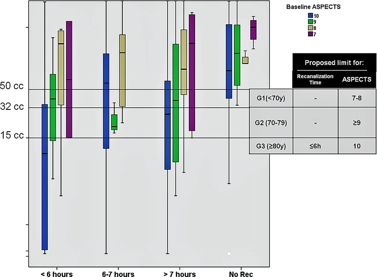
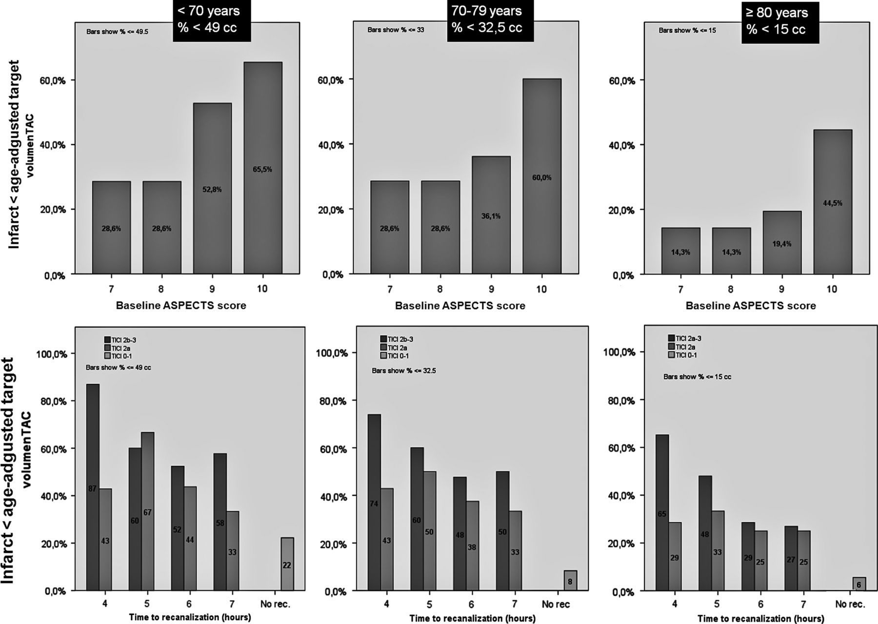
Probabilities of achieving an infarct volume less than the age-adjusted target decreased according to baseline ASPECTS, time and degree of recanalization (figures 2 and 3).

Infarct volume distribution according to time to recanalization and baseline Alberta Stroke Program Early CT score (ASPECTS). Age-adjusted thresholds for good outcome.

Percentage of patients achieving an infarct volume less than the age-adjusted target according to the Alberta Stroke Program Early CT score (ASPECTS) on initial CT and time/degree of recanalization.
Discussion
In addition to confirming the predictive power of age and infarct volume on further outcome, this study shows that the acceptable final lesion compatible with a good outcome varies and decreases according to patient age. Advancing age may be associated with reduced tolerance for infarct volume. While most young patients can afford a 50 mL final lesion and still be independent at 3 months, octogenarians need to show virtually no residual lesion (<15 mL) to achieve good functional recovery after endovascular treatment.
This difference may be due to a number of factors such as the coexistence of multiple comorbidities or an impaired capacity of neurorestoration in older patients.8 The adult brain retains the capacity for neurogenesis and neuroplasticity to recover after pathological processes such as stroke.11 However, this phenomenon diminishes with ageing, leading to disproportionate consequences in the aged brain.12
Previous studies have indicated that the final infarct volume may represent an accurate indicator of procedural success and therefore should be used in preference to recanalization scores as surrogate markers of outcome in reperfusion trials for acute stroke.2–4 Syed et al found that a final infarct volume cut-off of 40 mL best differentiated between favorable and unfavorable outcomes. This value falls in the middle of the age-adjusted infarct volume range described in our study. Our findings may have a practical application in the decision-making process when evaluating patients with acute stroke for endovascular procedures. Several classical factors such as age, symptom duration or severity were found to be associated with outcome in the univariate analysis. However, in the multivariate analysis the age-adjusted target infarct volume was found to be one of the strongest independent predictors of long-term functional recovery. Adjusting the maximal affordable infarct volume to the patient age may increase the probability of selecting patients who will benefit from endovascular therapies. In order to guide the decision-making process, we elaborated the probability charts to achieve the age-adjusted infarct volume according to relevant variables available on patient admission such as time from symptom onset or ASPECTS score on initial CT. According to these charts, endovascular procedures for patients aged >80 years may still have relatively good results, but only under more restrictive inclusion criteria than in younger age cohorts— that is, baseline ASPECTS score ≥9 and recanalization achieved ≤4.5 h from symptom onset.
We also calculated the chances of achieving an infarct volume less than the age-adjusted target according to time to reperfusion or degree of final recanalization. This information can also be used in the angio-suite when deciding to pursue recanalization efforts in longer time windows or seeking perfect angiographic results (TICI 2b–3) rather than partial recanalizaton. While in some cases younger patients may still attain the target infarct despite achieving partial TICI 2a recanalization, older patients will imperatively require successful (TICI 2b–3) and early (<4.5 h) recanalizaton.
The above considerations may also be used as a guide in the design of inclusion criteria for new endovascular therapy trials for acute ischemic stroke where age-adjusted final infarct volume may represent a valuable surrogate marker of favorable long-term outcome.
Theoretically, weighting the age-adjusted volume according to infarct location may increase its predictive power according to the strategic topographical stoke concept. Our study was designed to offer a reliable, rapid and replicable tool to predict outcome. Future studies may address the impact of the anatomical location of stroke on disability— that is, according to specific affected ASPECTS regions.
Using the follow-up CT scan to measure the final infarct volume instead of a diffusion-weighted MRI may represent a limitation in our study since MRI may offer a more accurate value of the real final volume. However, we preferred the 24–36 h CT scan because it was available in nearly all patients and using MRI values would represent a bias since it is frequently unavailable in patients with most severe strokes.
Conclusion
Age-adjusted infarct size might represent a powerful surrogate marker of stroke outcome and further refine the predictive accuracy of infarct volume on prognosis in patients with stroke undergoing endovascular treatment. This information may be useful in the design of new trials to individualize selection criteria for stratifying different age groups according to baseline ASPECTS score and time from symptom onset.
References
Footnotes
-
Contributors MRib, AF, EM and CAM participated in the conception and design of the study. MRib, AF, EM and JAS analyzed and interpreted the data. MRib, MRub, AT, PC, JP, DR-L and MM treated and included the patients in the study. MRib performed the statistical analysis and wrote the article. All authors reviewed and approved the manuscript.
-
Funding MRib is the recipient of a grant from Instituto de Salud Carlos III, Ministerio de Economía y Competitividad (FIS: PS09/01660).
-
Competing interests None.
-
Patient consent Obtained.
-
Ethics approval Ethics approval was obtained from the CEIC Institut de Recerca Vall d'Hebron.
-
Provenance and peer review Not commissioned; externally peer reviewed.