Article Text

Abstract
The syndrome of hemispatial neglect is characterised by reduced awareness of stimuli on one side of space, even though there may be no sensory loss. Although it is extremely common, it has proven to be a challenging condition to understand, and to treat. New insights from detailed behavioural and anatomical studies in patients, as well as functional imaging in healthy individuals, have begun to reveal some of the component deficits underlying the disorder. This review focuses on important clinical issues in neglect, including bedside diagnostic tests and emerging therapeutic and rehabilitation methods, involving both behavioural and drug treatments.
- anosognosia
- extinction
- neglect
- BIT, behavioural inattention test
- fMRI, functional magnetic resonance imaging
- SPECT, single photon emission computed tomography
- STG, superior temporal gyrus
Statistics from Altmetric.com
- BIT, behavioural inattention test
- fMRI, functional magnetic resonance imaging
- SPECT, single photon emission computed tomography
- STG, superior temporal gyrus
Hemispatial neglect is a common disabling condition following unilateral brain damage, particularly of the right hemisphere. Although it can be caused by various different pathological conditions, it is most often observed after cerebral infarction or haemorrhage and affects up to two thirds of right hemisphere stroke patients acutely.12 Patients with neglect often fail to be aware of or acknowledge items on their contralesional side (the left side for patients with right brain damage) and attend instead to items towards the same side as their brain damage—their ipsilesional side. Their neglect may be so profound that they are unaware of large objects, or even people, in extrapersonal space. Neglect may also extend or be confined to personal space, with patients failing to acknowledge their own contralesional body parts in daily life.3–5 Moreover, some patients fail to use their contralesional limbs even if they have little or no weakness—so called “motor neglect.”67 Importantly, many neglect patients may be unaware that they have any of these problems (anosognosia), sometimes denying that there is anything wrong with their perception or control of movement.78 Perhaps it is not surprising, therefore, to find that enduring neglect is a poor prognostic indicator for functional independence following stroke.9–13
In this selective review, we focus on some important clinical aspects of the neglect syndrome. First, we consider how bedside examination may best reveal and quantify the degree of neglect. Second, we discuss the potential mechanisms underlying neglect. Most contemporary views of the neglect syndrome consider it to be a heterogeneous condition14–26 consistent with the heterogeneous nature of the associated lesion sites152728(see Vallar29 for one proposed taxonomy). Here, we argue that neglect emerges as a result of a combination of component cognitive deficits that may vary across patients and need not be neglect specific.1824 Finally, we discuss treatments, focusing particularly on recent research using prism therapy,3031 which has shown promising beneficial effects and may well have an impact on clinical practice.
BEDSIDE EXAMINATION
Does the patient have neglect?
Patients with the most severe unilateral neglect are obvious “from the end of the bed”—that is, the diagnosis may be made by simple observation from a distance. The patient with a large infarct in the right middle cerebral artery territory may have their head and eyes turned to the extreme right and never gaze to the left. When presented with food or a newspaper to read, they may show interest in items only to their right, ignoring those to their left. Similarly, when approached by ward staff from their left they may fail to acknowledge them or, if they are spoken to, they may orientate themselves to the right and reply with their gaze directed away from the person they are addressing. Note that such behaviour would be very unusual for a hemianopic patient who does not suffer from neglect as well, although, as we discuss below, the distinction between pure neglect and neglect plus hemianopia is not always straightforward.
Most patients with neglect are not necessarily so easy to identify as this. Moreover, it is increasingly becoming important to measure the severity of neglect. Just like more conventional neurological examination measures, such as the degree of limb weakness, this helps the clinician to track the progress of the patient. Many patients with neglect following stroke improve within a few weeks, but some continue to show persistent neglect and it is these individuals who are likely to require rehabilitation input. Early identification of patients who show little or no sign of improvement may facilitate their referral to specialised rehabilitation units or identify those who may need more intensive occupational therapy or physiotherapy.
Several simple bedside screening tests have been developed for the assessment of neglect. Although many clinicians are familiar with object copying (fig 1A) and clock drawing tests (fig 1B), these are not very sensitive on their own,32 and are also not always easy to score in a graded manner. Furthermore, patients with constructional apraxia may perform poorly on such tasks, showing copying errors on both the left and the right side of space, even though they do not show any spatial neglect. Fortunately, there are various other relatively simple tests available that are often used by neuropsychologists and therapists assessing patients on the ward, although perhaps not so familiar to many practising neurologists or physicians. Batteries of such tests have been developed,33–36 largely because no single test alone is able to detect neglect in all patients.323537 Moreover, there are many reports of clear dissociations, with some patients showing neglect on certain tasks but not on others.353839 Here we focus on only a few of the available tests, concentrating on some of the common ones that are used at the bedside.

Typically, right hemisphere patients with left neglect omit elements to their left when copying simple objects (A), drawing a clock face (B), and cancelling targets among distractors (C). They also tend to err to the right when asked to bisect a horizontal line (D). When asked to name objects in their surroundings, they will tend to name only those on the right. Crosses in (E) mark the locations of reported objects with respect to the patient.
One of the most useful types of test for neglect is the cancellation task. There are several different versions available; all of them require the patient to find and cancel (mark with a pen) target items distributed on an A4 sized sheet of paper placed directly in front of them. Some cancellation tasks have only target items—for example, the Albert’s task40 or line cancellation from the behavioural inattention test (BIT) battery,33 but most of them have targets embedded within an array of many different types of distractor items—for example, the bells test,41 star cancellation from the BIT,33 and the Mesulam shape cancellation test.42 Many right hemisphere patients with left neglect cancel items on only the right side of cancellation tasks, omitting targets to the left (fig 1). An important clue to the presence of neglect is that most such patients also start to search from the right of the array,32 whereas most control subjects who read left to right start on the left. In our experience, dense cancellation tasks with distractors are usually better and more sensitive in detecting neglect than the simpler cancellation tasks that have no distractor items (see also Halligan et al37). Moreover, they detect neglect more frequently than any other single test,353739 although there are a few patients who perform well on cancellation but show neglect on other tasks43—hence the need to use more than one screening measure for neglect.
Another simple pen and paper task that has been extensively used is line bisection. A long horizontal line marked on an A4 sheet of paper is placed in front of the patient, who is asked to mark the apparent midpoint of the line. Many right hemisphere patients with left neglect, particularly those with posterior lesions,3238 tend to mark the apparent middle of the line well to the right of the true midline (fig 1D). Patients with a left hemianopia but without neglect tend to bisect the line slightly to the left, perhaps because they are aware of their visual deficit and attempt to compensate for it.44 A similar contralesional bias in performance can also be seen in left neglect patients if the line to be bisected is small (the so called “crossover” effect45) but in normal clinical practice large horizontal lines (18–20 cm long) are normally used, so this is not a confounding factor.
Asking the patient to report 10 objects in the room around them provides another rapid and useful measure.146 Provided that the patient is not situated to the extreme left or right of a room, this test may reveal a spatial bias, with neglect patients often reporting items only or mostly to their ipsilesional side (fig 1E), whereas patients with pure hemianopia will tend to compensate by moving their head and eyes towards contralesional space. Moreover, patients with pure hemianopia are typically aware of their deficit.
All the tests that we have discussed so far depend heavily on vision or visuomotor control. Some patients with neglect may perform normally on such tasks, but show personal neglect (ignoring the contralesional side of their body) or motor neglect (failing to use their contralesional limbs despite the fact that strength in these may be intact or only mildly reduced). Personal neglect may be detected formally by asking the patient to gesture how they would groom themselves—for example, comb their hair, shave, or put on make up.547 More often, however, such neglect is observed by carers. In our experience motor neglect67 is also most often detected by therapists or carers, who remark on the lack of use of a contralesional limb even though it is strong. Finally, many patients with neglect also show lateralised spatial deficits on tests of representational neglect—for example, if they are asked to recall a familiar scene from memory, right hemisphere neglect patients may ignore the left side,4849 and similarly with clock drawing from memory.
In the clinical setting, if the available time is limited to a brief assessment, the combination of a dense cancellation task with clock drawing and figure copying may be sufficient to pick up over 70% of neglect patients.32 However, if more time is available, behavioural assessments of neglect in daily life usually are more sensitive.3235
Does the patient suffer from hemianopia as well as neglect?
Many neglect patients suffer from hemianopia as well as neglect.50 In addition, there are also two other types of patient: first, there are individuals who suffer from hemianopia but do not show neglect; second, there are patients who show neglect on bedside testing or on clinical observation but who nevertheless have full visual fields to confrontation.51 It is usually operationally straightforward to confirm the presence of neglect by using the tests we describe above. In the absence of neglect, the presence of a contralesional field defect which shows a strict demarcation at the vertical meridian in both eyes is referred to as homonymous hemianopia. The real problem is deciding whether a failure by a patient to report a contralesional stimulus is due to neglect alone, or neglect plus hemianopia. Disentangling “absolute” field defects from neglect is not always easy and some question the validity of making such a distinction.52
The fact that two distinct syndromes may co-occur within the same patient is perhaps best illustrated by individuals who have complete loss of vision in the lower contralesional quadrant (regardless of the size or illumination of the test stimulus), but who nevertheless can report a salient stimulus in the upper quadrant and also show neglect on standard tests. We consider such a patient to have an absolute sensory defect (manifest as an inferior quadrantanopia), plus neglect. In our experience, assessment of such field defects is best done by careful clinical examination at the bedside rather than by the use of automated perimetry, which tends to overestimate the apparent “absolute” field defect.53 If there is evidence of a field defect on initial testing with small targets (for example, a hatpin) we repeat testing with larger targets (such as fingers) before we are fully satisfied of an “absolute” field defect. Even under these circumstances, we agree that it is sometimes difficult to distinguish clinically between dense neglect and neglect plus hemianopia.52
Markers of neural activity evoked by a visual stimulus that the patient fails to be aware of may be a useful way of distinguishing between neglect and absolute sensory loss. Thus evoked responses and fMRI have both been used to show that stimuli which fail to be reported by a patient may nevertheless produce brain responses.54–56 Moreover, behavioural studies have demonstrated that such stimuli may be (implicitly) processed to relatively high levels.5758
Does the patient have extinction?
If a patient has full visual fields but nevertheless fails to report the contralesional stimulus when it is presented simultaneously with an ipsilesional stimulus, he is said to show “extinction.”5960 Many patients have extinction but not neglect on standard tests, and some consider extinction to be a mild type of neglect or a sign of “inattention.” In addition, many patients with neglect also show extinction, which may be considered one component of the neglect syndrome, although we emphasise that it may not be present in all neglect patients. If the patient appears to have a field defect in addition to neglect, it may still be possible to detect extinction by presenting two stimuli in the intact field.61 Finally, it is also worth noting that extinction may also occur in other modes—for example, tactile and auditory—as well as being cross modal, as when a visual stimulus to the right “extinguishes” a tactile stimulus on the left.62 The significance of extinction for understanding the possible competitive attentional mechanisms underlying neglect is discussed below.
Is the patient anosognosic?
Although patients with severe anosognosia are often identified through conversation at the bedside, many may not reveal unawareness of one or more of their neurological deficits so easily. There are various relatively simple structured instruments863 that can be helpful in screening for such deficits, which may have an important impact on functional recovery or rehabilitation potential. Neglect, as defined by the bedside tests we have discussed, may occur without anosognosia and vice versa, but many patients suffer from both conditions.763
MECHANISMS UNDERLYING NEGLECT
Damage to many different brain regions causes neglect
Lesions of the right hemisphere are far more likely to lead to severe and enduring neglect than left hemisphere damage,264 perhaps because of the specialisation of the latter for language. Cortical damage involving the right inferior parietal lobe or nearby temporo-parietal junction has classically been implicated in causing neglect.50 It has become apparent, however, that the syndrome may also follow focal lesions of the inferior frontal lobe2865(fig 2), although lesions confined to the frontal lobe may lead to a more transient neglect (see, for example, the case described by Walker et al66). More commonly, however, large middle cerebral artery strokes span both the critical parietal and frontal regions, resulting in a severe and persistent neglect syndrome that has a profound impact on the daily behaviour of patients.

Cortical right hemisphere brain regions that have been associated with neglect include the angular (ang) and supramarginal (smg) gyri of the inferior parietal lobe (IPL), the temporo-parietal junction (TPJ), the superior temporal gyrus (STG), and the inferior (IFG) and middle frontal (MFG) gyri.
Recently, a provocative anatomical study has challenged the conventional view that inferior parietal lobe or temporo-parietal junction lesions are the critical posterior cortical locations associated with neglect. Karnath and colleagues67 have instead proposed that the key region that must be damaged is the mid-superior temporal gyrus (STG). However, a subsequent investigation, using higher resolution lesion mapping methods, showed that, although the STG may well be involved in many neglect patients (50% in the sample), damage to this region is not invariably associated with the condition.68 Rather, the critical brain region involved in every case of neglect following middle cerebral artery stroke was found to be the angular gyrus of the parietal lobe.
In addition to cortical damage, subcortical ischaemic lesions in the territory of the middle cerebral artery involving the right basal ganglia or thalamus may also produce neglect,2869 but this may reflect diaschisis or hypoperfusion in overlying parietal and frontal regions, as demonstrated by both SPECT and magnetic resonance perfusion.7071 Finally, some patients with posterior cerebral artery territory stroke also suffer from neglect, although these individuals have been less well studied. Some groups have observed that while small lesions involving the occipital lobe lead to hemianopia, larger strokes extending into the medial temporal lobe also lead to neglect.506872 Specifically, Mort et al have recently shown68 that the key medial temporal area that is damaged in these patients is the parahippocampal region, an area that has strong connections with the parietal cortex,7374 and may be considered an important gateway for parietal information to the hippocampus. Although lesions of the right parahippocampal region are traditionally associated with topographical disorientation, there are reports of patients who also show neglect.75 What remains to be determined is whether neglect following extensive posterior cerebral artery infarction is in fact caused by diaschisis in the parietal cortex or is a separate disorder distinguished by unique underlying component deficits.
Many different mechanisms may contribute to neglect
Given the variety and widespread nature of the lesions—both cortical and subcortical—that have been implicated in neglect, it is perhaps not surprising that many different mechanisms are now considered to contribute to the syndrome. Moreover, functional imaging studies in healthy individuals have shown that many different functions might be subserved by subregions within even the inferior parietal lobe.18242676 Increasingly, neglect is considered to consist of a number of component deficits, with the precise combination varying from patient to patient, and presumably determined by the exact location and extent of brain damage. A second critical concept that is emerging is that the mechanisms underlying neglect need not be neglect specific: they may occur separately on their own in patients without neglect.1824 When combined with other component deficits, however, they may lead to neglect. These perspectives have important implications not only for understanding the neglect syndrome but also for treating it.
Because of space constraints, it is not possible here to detail all the component deficits that have been considered to play a role in neglect or related disorders such as anosognosia.714–2023–2577 Rather, our objective here is to provide a brief overview and use a few examples to illustrate the key concepts that we have outlined above.
Various important spatially lateralised component deficits have been proposed to underlie neglect. A disorder of directing attention to the left is considered to be a “core” problem by many investigators. It is debatable whether this reflects an intrinsic graded bias to direct attention rightwards following right hemisphere damage7879 or because items on the right invariably “win” over objects to the left in the competition for selection, as some have argued to be the case in extinction8081; or because of difficulty in disengaging attention and shifting it leftward,198283 as others claim also for extinction. Several investigators have also raised the possibility that neglect may result from an impaired representation of space,4884 which can be in multiple frames of reference (for example, retinotopic, head centred, trunk centred) or be specific to near or far space.85–87 Still others have considered that neglect may also reflect a directional motor impairment, with patients experiencing difficulty in initiating or programming contralesional eye or limb movements.8889 Of course, these proposed lateralised component deficits are not mutually incompatible90 and several may coexist within the same individual—for example, directional motor and attentional deficits have been shown to be present in both parietal and frontal neglect patients.91
In addition to these directional deficits, it is increasingly becoming apparent that non-spatially lateralised mechanisms may also contribute to neglect.1824 For example, impairments in sustained attention,92 selective attention at central fixation93 or in both visual fields,9495 a bias to local features in the visual scene,229697 as well as a deficit in spatial working memory98—even within a vertical array99—have all been implicated in the neglect syndrome. Importantly, none of these deficits has traditionally been considered to be neglect specific. Instead, they have been viewed as coexisting deficits, as they may occur independently in patients without neglect—that is, they are “doubly dissociable” from the neglect syndrome. However, several investigators argue that when such non-spatially lateralised deficits combine with spatially lateralised ones, they exacerbate any directional deficit and thereby have a significant impact on the neglect syndrome, reducing the potential for recovery.1824100
Such a view of neglect has two important consequences. First, it brings to bear insights from other branches of cognitive neuroscience—such as spatial working memory and sustained attention—that have hitherto not been considered to be important for understanding neglect. Second, it raises the possibility of targeting treatments towards specific component deficits that may not be neglect specific but nevertheless are important in determining the severity of neglect. The full potential for such treatments has yet to be tested, but recent work suggests this may be a promising avenue in the near future.
TREATMENT AND REHABILITATION
Scanning therapy and hemianopic patching
Initial attempts to rehabilitate neglect often attempted to encourage patients to direct their gaze towards contralesional space,101–103 and functional imaging has suggested this may be associated with increased activation of intact right hemisphere regions that are involved in visual search.104 Although these approaches showed some success in reducing neglect within a particular task (for example, in reading by cueing patients to find a red line marked by the investigators on the left margin102), patients showed little or no generalisation of their improved scanning behaviour to tasks outside of the training environment.12 This failure to generalise may partly be attributable to the dependence of these paradigms on the patients being aware of their deficit and deliberately modifying their behaviour (“top-down”) as a consequence. Unfortunately, as many patients with neglect are often unaware of their deficit, they may require frequent reminders to scan left, and in complex real world environments, cues (such as the red line used to improve reading of words on the left) are not readily available.
A recent alternative approach consists of using spectacles that occlude the good (ipsilesional) side of vision in each eye, effectively forcing neglect patients to direct their gaze to their contralesional side,105 whatever the visual environment. Although such “hemianopic patching” seems promising, the reported benefits have been modest,106 perhaps because patients who might benefit need to be selected carefully. Many patients do not tolerate these spectacles well, presumably because their natural inclination is to gaze towards the now occluded ipsilesional visual field, and, in our limited experience with this technique, compliance is not optimal.
Inducing shifts in spatial representations
Several groups have attempted to involve a more direct approach to altering the impaired representation of space in neglect. The methods they have used include caloric, or vestibular, stimulation,107108 contralesional limb activation,109 trunk rotation,110 neck muscle vibration,111112 and electrical stimulation of the neck.113 Although the mechanisms involved in these different techniques vary they have all been shown to produce an improvement in some aspects of neglect. Furthermore, they all produce an automatic (“bottom-up”) change in behaviour, or recalibration of the sensorimotor mechanisms recruited, that does not depend upon patients adopting (“top-down”) a new control strategy to look leftwards. Perhaps as a result, improvements in performance have been shown—at least in some cases—to generalise to tasks that were not used in training.
Rubens107 was the first to demonstrate the potential of these techniques using caloric stimulation, which involves the application of cold water to the contralesional ear (or warm water to the ipsilesional ear), causing a vestibular induced contralesional shift in gaze. This produces a transient amelioration in the patient’s neglect during and after application (for 10 to 15 minutes) across a range of tasks. However, while this technique is of theoretical interest, the short duration of its effects, together with the discomfort of application, renders it impractical as a basis for rehabilitation.
Because of the possible role of an impaired representation of space anchored to the midline of the trunk, Karnath and colleagues proposed that shifting the perceived location of the body midline into the contralesional field might also ameliorate neglect.110112114 They found that if the orientation of a patient’s trunk was rotated leftwards, while they kept their head and eyes fixed straight ahead, performance for stimuli on the left improved significantly.110 A similar effect after vibration of the contralesional neck muscles—which produces the same proprioceptive feedback from the neck muscles as a contralesional trunk rotation—has also been reported.114 Moreover, when patients were treated with neck muscle vibration in combination with scanning training, a long lasting improvement (discernible even after two months) was observed on visuomotor tasks that had not been used in initial training.112
Robertson and colleagues have found that active movements of part of the contralesional half of a patient’s body (a finger) can produce improvements on a number of tests of neglect,109115116 particularly in near compared to far space115(see also Frassinetti et al117). Although this spatiomotor cueing technique has also been shown to be effective in patients with contralesional limb weakness,117 the prevalence of severe hemiparesis and sensory loss in neglect patients may limit the number of individuals who might benefit from this technique. Nevertheless, one trial has shown that such treatment may reduce the length of hospital stay in patients with neglect significantly.118
Prism adaptation
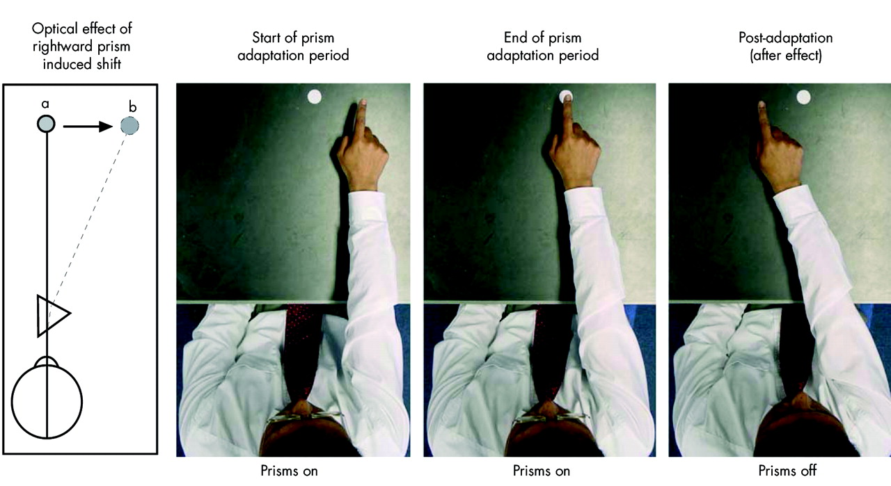
A new type of treatment which is cheap, simple to apply, apparently free of side effects, and which generalises across a range of tasks for many weeks afterwards has attracted a great deal of interest recently. The benefits of prism adaptation were first reported by Rossetti et al,30 who examined the effects of adaptation to a 10° rightward horizontal displacement of their visual field by prisms in 12 neglect patients. While wearing the prisms the patients repeatedly pointed (for only 50 trials) to targets 10 either side of their body midline (but optically lying either straight ahead or 20° to the right (fig 3)). Immediately after adaptation they found that neglect was ameliorated across all five of the neglect tests they used; this improvement was even greater after a further two hours. A control group of neglect patients that underwent exactly the same procedure but wearing flat lenses showed no significant improvement in their performance.
Adaptation to a rightward displacement in an observer’s vision produced by a prism. When viewing a scene through the wedge prism, all points are displaced horizontally to the right with respect to the optical axis of the retina (first panel). Hence, an object at point “a” will appear to be located at point “b”. The adaptation process requires the observer to reach for targets repeatedly within the visual scene. At the start of the process (second panel), participants will misreach to the right of the target, an error referred to as the direct effect. The error will swiftly diminish and disappear entirely as the participant adapts to the visual shift (third panel). However, to enable the participant to adapt fully, approximately 50 repetitions should be completed. When the prisms are removed the participants will misreach in the opposite direction to the visual shift (fourth panel), an error referred to as the after effect. In normal observers this after effect will disappear after only a few minutes.
Frassinetti et al further demonstrated the therapeutic potential of prism adaptation by showing that it can result in a long term amelioration of neglect.31 Patients in their study were given brief prism adaptation twice a day for two weeks and their performance was compared with a matched control group who underwent a standard rehabilitation programme. The participants were tested on a wide range of neglect assessments, ecologically valid real world tasks, and at different spatial frames (personal, near, and far space). Nearly all the patients who had prism treatment showed a significant improvement in neglect after the first session in virtually every task. Remarkably, this improvement increased in magnitude each time the patients were assessed up to, and including, five weeks after the last session of adaptation. Other studies have also shown that prism adaptation is associated with improvements in representational neglect,119120 neglect dyslexia,121 postural imbalance in hemiparesis,122 haptic neglect,123 and tactile extinction.124 Additionally, McIntosh et al reported that the benefits of prism adaptation can extend to a chronic neglect patient treated nine months after her stroke.123
The mechanisms underlying the effectiveness of prism adaptation are not yet precisely understood. However, the general conclusion of the previous studies is that the results cannot be explained merely by a leftward motoric bias of the right arm during adaptation to prisms. First, improvements occur in domains that do not require a limb specific motor response—for example, reporting objects around a room or representational neglect.31119120 Second, there is no correlation between the duration of the improvement in performance in tests of neglect and the duration of prism after effects, measured by pointing straight ahead without visual feedback.31 Thus most studies have concluded that prism adaptation affects higher level spatial representations that are disrupted in neglect.3031 Rossetti et al claimed that the stimulation of low level neural mechanisms that monitor and correct errors between the actual and expected positions of the arm in prism adaptation might correct the biases introduced by neglect.30 However, as Frassinetti et al note,31 an alternative explanation is that the improvements in neglect reflect changes in the control of the oculomotor system. Amelioration of neglect has been shown after interventions that cause an involuntary shift of gaze into the neglected field, for example vestibular stimulation.107 Additionally, previous studies of prism adaptation in normal subjects have reported appreciable oculomotor shifts.125
This raises the general issue of the appropriate measurement of the effects of adaptation. To our knowledge all studies of prism adaptation and neglect have measured adaptation by comparing pointing along the body midline before and after the application of prisms using the adapted arm. However, this does not measure the total effects of adaptation; nor does it, as has been suggested,31126 measure a shift in the perceived body midline but merely the adaptive shift within the head–arm system.125 To assess the effects of adaptation on the sagittal body axis it is necessary to use a measure that is independent of any direct motoric adaptation. Unfortunately, the prevalence of hemiparesis in neglect means that the obvious solution of using the arm not employed in the adaptation task would not be realistic for the majority of patients. However, the adaptive after effect in the oculomotor system can be readily measured by setting the position of a line on a computer monitor so that it appears to lie directly ahead.125 An important question for future research directed towards understanding why prisms are effective would be to examine the magnitude and persistence of this after effect, and to assess how it correlates with improved performance by neglect patients. New data have begun to suggest that the effectiveness of prism therapy is not due to altering the spatially lateralised gradient of attention, at least in patients with mild neglect.127
Treating non-spatially lateralised deficits
Is it possible to ameliorate the severity of the lateralised deficit in neglect using treatments that target non-spatially lateralised impairments (that is, those that affect both sides of space)? Robertson and his colleagues tested this hypothesis directly by investigating whether increasing patients’ alertness would lead to an improvement in their spatial bias.128 They examined thresholds for detecting whether a stimulus on the left preceded or followed the appearance of a comparable object on the right. On this task, neglect patients showed a strong spatial bias, typically judging the appearance of both stimuli to be simultaneous when the left object preceded the right one by half a second. Remarkably, this spatial bias was abolished if the stimuli were preceded by a short tone, attributed to a boost in the patients’ alertness because the tone did not contain any information that would predict which object would come first. Furthermore, the effect occurred even when the tone originated on the right which, if anything, would tend to cue the patient away from the neglected field.
A behavioural technique—more appropriate for treating sustained attention deficits in clinical settings—has also been developed.129 Neglect patients were required to carry out a variety of tasks that required sustained attention, for example sorting coins or cards. While carrying out the task the experimenter would intermittently prompt them verbally to attend. Patients were then gradually trained to prompt themselves subvocally. They were also made aware of their sustained attentional deficit difficulties and the importance of this self alerting process. The eight patients showed considerable improvements, 24 hours after training, in tests of sustained attention and spatial neglect. However, the nature of the intervention requires patients to be aware of their deficit, as well as the situations in which it is necessary to alert themselves. The degree to which patients are able to do this may limit the general applicability of this technique.
An alternative to behavioural approaches for the treatment of non-lateralised cognitive deficits associated with neglect might be the use of targeted pharmacological interventions. Specifically, it has been suggested that impaired sustained attention could be targeted either through noradrenergic drugs known to modulate vigilance levels in healthy volunteers,130 or through cholinergic compounds—for example, acetylcholinesterase inhibitors—that are currently used to improve cognitive function in several conditions including Alzheimer’s disease and vascular dementia.131132
By contrast, spatial working memory deficits across saccades98 might be targeted using dopaminergic drugs. Physiological evidence from studies in rhesus monkeys suggests that memory for the locations of saccadic targets are modulated by dopamine D1 receptor agents.133 Previous studies using the dopamine agonist bromocriptine have reported both positive and negative results.134–136 Such conflicting findings may partly reflect the heterogeneity of patients included in the studies as well as the fact that bromocriptine acts mainly on D2 dopamine receptors. A future study might profitably examine the effects of an agent that primarily targets D1 receptors in selected neglect patients who have been shown to have a spatial working memory deficit.
CONCLUSIONS
In this review, we have focused on clinical aspects of neglect. Recent findings emphasise that the neglect syndrome is a heterogeneous condition with different combinations of deficit occurring in different patients. While some of these components are spatially lateralised, others are not. Treatments for neglect are unlikely to be successful unless they are tailored to the underlying cognitive deficits in individual patients. Promising new developments using behavioural and drug interventions have begun to offer some new hope for this common debilitating condition.
Acknowledgments
We are extremely grateful to the many patients who have participated in our research, including those from the stroke units at Charing Cross Hospital and St Thomas’ Hospital and the acute brain injury unit, National Hospital for Neurology and Neurosurgery, London. We would also like to thank both reviewers for their helpful comments and suggestions. This work is funded by the Wellcome Trust.
REFERENCES
Footnotes
-
Competing interests: none declared