Article Text

Abstract
Background IgG anti-GQ1b antibodies are associated with Fisher syndrome (FS), Bickerstaff brainstem encephalitis (BBE), acute ophthalmoparesis and overlap of FS or BBE with Guillain–Barré syndrome (GBS) (FS/GBS or BBE/GBS). It has not been clearly established if the primary pathology of these disorders is demyelinating or axonal in nature. Rapid resolution of conduction slowing or block without signs of demyelination–remyelination has been reported in axonal subtypes of GBS that are associated with IgG anti-GM1 or -GD1a antibodies. We hypothesised that such reversible conduction failure would be also observed in FS and related disorders.
Methods Serial nerve conduction studies were prospectively performed in 15 patients with FS and related conditions.
Results Neither conduction block nor abnormal temporal dispersion was observed in any of the nerves at any point in all the patients. Conduction velocities for none of the nerves were in the demyelinating range. The amplitude of sensory nerve action potential was decreased in three FS, one FS/GBS and two BBE/GBS patients. Compound muscle action potential amplitudes were decreased in the two BBE/GBS patients. These decreases in amplitudes of sensory nerve action potential and compound muscle action potential promptly resolved without significant change in duration on serial studies.
Conclusions Reversible conduction failure was seen in six of the 15 patients with FS and related disorders on serial nerve conduction studies. There were no signs of demyelination or remyelination in the 15 patients. The pathology appears to be primarily non-demyelinating. We believe these conditions form a continuous spectrum with axonal GBS.
- Guillain–Barré syndrome
- Fisher syndrome
- anti-GQ1b antibody
- neurophysiology
- ganglioside
- CIDP
- Miller Fisher syndrome
Statistics from Altmetric.com
- Guillain–Barré syndrome
- Fisher syndrome
- anti-GQ1b antibody
- neurophysiology
- ganglioside
- CIDP
- Miller Fisher syndrome
IgG autoantibodies against ganglioside GM1 or GD1a are associated with reversible conduction failure, characterised by rapid resolution of conduction slowing and block without signs of remyelination such as temporal dispersion and decreasing conduction velocity, as well as axonal degeneration.1 These are the electrophysiological hallmarks of acute motor axonal neuropathy (AMAN), an axonal subtype of Guillain–Barré syndrome (GBS). In an animal model of AMAN, produced by sensitising rabbits with GM1, IgG antibodies bind to nodes and paranodes and activate complement, resulting in sodium channel cluster disappearance and paranodal myelin detachment in anterior spinal nerve roots.2 Paranodal changes, along with the nodal lengthening, have also been demonstrated in AMAN patients.3 These pathological changes can induce conduction failure and muscle weakness. A recent study has shown that the IgG monoclonal anti-GD1a antibody, injected into rat sciatic nerves, caused the deposition of IgG and complement products on the nodal axolemma and disrupted clusters of nodal and paranodal molecules predominantly in motor nerves, and induced reversible conduction block.4 These electrophysiological and immunopathological findings indicate that IgG anti-GM1 or -GD1a antibodies could induce reversible conduction failure in AMAN.
Fisher syndrome (FS) characterised by ophthalmoplegia, ataxia and areflexia has been well established as a subtype of GBS. Some FS patients develop weakness and clinical features that overlap with GBS (FS/GBS).5 Since the identification of IgG anti-GQ1b antibodies in FS,6 remarkable progression has been made in our understanding of FS and related conditions. Bickerstaff brainstem encephalitis (BBE) and FS share many clinical features and the association with anti-GQ1b antibodies, suggesting a common autoimmune aetiology.7 These autoantibodies are also associated with incomplete forms of FS, namely, acute ophthalmoparesis (AO) (without ataxia) and acute ataxic neuropathy (without ataxia).8 ,9 ‘Anti-GQ1b antibody syndrome’ includes these conditions and related ones such as FS/GBS and BBE overlapping with GBS (BBE/GBS).10 Recognition of the relationship between these various anti-GQ1b antibody-associated variants of GBS is likely to yield useful clues to the pathophysiology of these subtypes of GBS.
It has yet to be established if the primary pathology of FS is demyelinating or axonal in nature. One approach to study this would be to analyse FS as well as its related conditions. Both FS and AMAN are more closely linked to the presence of anti-ganglioside antibodies and preceding Campylobacter jejuni infection than acute inflammatory demyelinating polyneuropathy.11 ,12 AMAN and FS are also more common in non-Western countries.1 ,5 ,11–14 These observations raise the possibility of a closer relationship of FS to AMAN rather than acute inflammatory demyelinating polyneuropathy. There are only a few pathological studies of FS/GBS, BBE, BBE/GBS and acute ataxic neuropathy, and they lack sensitive tests such as teased fibre examinations to accurately differentiate demyelination from axonal pathology.15–18
As mentioned above, patients with AMAN develop weakness due to either axonal degeneration or reversible conduction failure. Resolution of conduction failure and conduction block without signs of demyelination–remyelination in convalescent-stage nerve conduction studies explains the quick recovery in several AMAN patients.1 Sequential electrodiagnostic studies are useful to delineate this reversible conduction failure of axonopathy and distinguish it from primary demyelination seen in other forms of GBS.19 In this study, we hypothesised that FS and related conditions would show similar electrophysiological changes on serial nerve conduction studies.
Methods
Patients and diagnostic criteria
A prospective evaluation of patients with FS and related disorders started at the National Neuroscience Institute in Singapore, August 2010. The institutional review board approved the study. We received written informed patient consent to perform this study. FS was defined by acute ophthalmoplegia, ataxia and areflexia or hyporeflexia with alert consciousness.20 Limb strength should remain within grade 4 or more on the Medical Research Council scale for muscle strength throughout the illness. BBE was diagnosed in the presence of ophthalmoplegia, ataxia and impaired consciousness. FS or BBE patients with limb weakness of Medical Research Council grade 3 or less were defined as FS/GBS or BBE/GBS.10 In AO, the extraocular muscle weakness should not be accompanied by ataxia, limb weakness or altered consciousness.9 Supportive evidence for the above conditions would be the presence of antecedent illness, serum anti-GQ1b antibodies and cerebrospinal fluid albuminocytological dissociation as well as the absence of alternative causes such as Wernicke's encephalopathy or brainstem stroke.20
Nerve conduction studies
Nerve conduction studies of at least one upper and one lower limb were performed using conventional methods. The contralateral limb was examined only to verify and compare abnormal results. The first nerve conduction study was done within 1 week from the onset of neurological symptoms. The second study was done between 1 and 5 weeks from the onset. If dynamic changes were noted, further studies were performed over the next few months until the nerve conduction parameters were stable.
Compound muscle action potentials (CMAP) and F-waves were recorded from the median, ulnar, tibial and peroneal nerves; and sensory nerve action potentials (SNAP) from median, ulnar and sural nerves using Nicolet VIKING IV electromyography machine (Carefusion, Middleton, Wisconsin, USA). Sensory nerves were stimulated antidromically. For motor nerve conduction studies, the filter was set at 2 Hz to 10 kHz. The corresponding values for sensory studies were 20 Hz to 3 KHz. Stimulus intensity and duration were optimised to ensure supramaximal stimulation. Skin temperature was maintained at or above 31°C. CMAP amplitude, duration and SNAP amplitude were measured using the initial negative phase. Normal values for the nerve conduction studies have been established previously on a cohort of 245 controls recruited from the same population. On sequential studies, a change in SNAP amplitude of 45% for median nerve, 49% for ulnar nerve and 60% for sural nerve was considered significant.21 Nerves were classified as either demyelinating or axonal according to previously established criteria.13 ,14 Conduction block was defined as 50% or more reduction in the proximal CMAP compared with the distal CMAP with temporal dispersion, defined as the increase in proximal CMAP duration divided by the distal duration, of <30%.22 Reversible distal conduction failure was defined as a decrease in distal CMAP amplitude that resolves without abnormal temporal dispersion (duration increase >30%) or other demyelinating features.1 Any conduction block observed would be categorised as demyelinating or axonal based on the follow-up studies. If the conduction block resolves with signs of demyelination or remyelination such as temporal dispersion and decreasing conduction velocity, then this conduction block would be labelled demyelinating type. Conversely, if follow-up studies show no temporal dispersion and conduction slowing, the conduction block would be attributed to reversible conduction failure and hence axonal in nature.
Results
Table 1 shows clinical and laboratory features of 15 patients studied between August 2010 and April 2012. Although this was a prospective series, we could not fix the interval between the studies for logistical reasons (table 2).
Clinical and laboratory findings
Serial nerve conduction studies
Fisher syndrome
Among the 10 FS patients, patients number 4, 5, 7, 9 and 10 had sensory symptoms or signs. Patients 3 and 5 respectively had a previous FS episode 10 and 4 years ago. All FS patients made full recovery over a period of 1–3 months. IgG anti-GQ1b antibodies were positive in all except Patient 4.
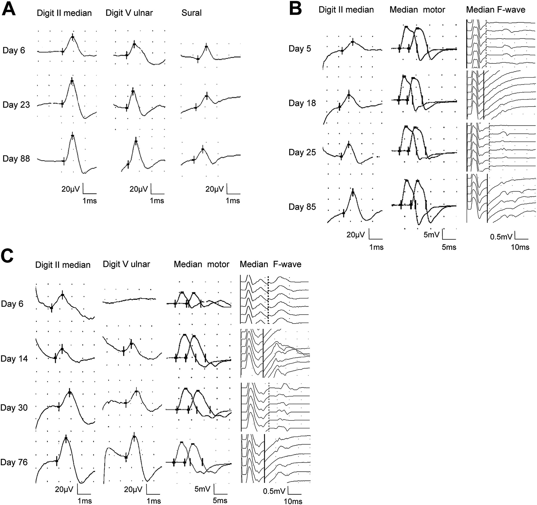
Figure 1A illustrates the serial, sensory studies of median nerve of Patient 5. She had normal median nerve and ulnar SNAP of 30 and 22 μV initially. Over the next few weeks, the median SNAP increased to 45 μV and the ulnar SNAP to 31 μV and then to 42 μV. The sensory conduction velocity of the median nerve remained stable at 43, 43 and 42 m/s. The corresponding sensory velocities of the ulnar nerve, respectively, were 58, 58 and 53 m/s. No significant change in SNAP duration was noted. The sural SNAP and motor conduction studies remained normal throughout without significant change.
Serial nerve conduction recordings. Serial recordings of sensory nerve action potentials (SNAP), superimposed median compound muscle action potentials (CMAP) from stimulation at wrist and elbow and F-waves. Patient 5 with Fisher syndrome (A) and Patient 11 with Fisher syndrome overlapped by Guillain–Barré syndrome (B) showed initial normal upper limb SNAP amplitudes that increase during convalescence. No abnormal dispersion of SNAPs was noted. Patient 12 with Bickerstaff brainstem encephalitis overlapped by Guillain–Barré syndrome (C) showed increase in upper limb SNAP and median CMAP amplitudes on serial examinations. No temporal dispersion was noted. The absent median F-waves reappeared with normal latency. Distal motor latency and conduction velocity were normal throughout.
Patient 6 had a similar increase in median SNAP (21, 30 and 35 μV) without change in conduction velocity (41, 44 and 46 m/s). The median distal motor latency (DML) was mildly increased. It improved significantly over the next 4 weeks. The residual changes were consistent with pre-existent, but asymptomatic, carpal tunnel syndrome. The improvement in DML was associated with no signs of remyelination such as abnormal temporal dispersion of the distal median CMAP. There were no other changes in the serial motor conduction studies. Patient 10 had similar abnormalities, but the median DML normalised completely in convalescent studies. The median SNAP amplitude of Patient 9, which was borderline abnormal, normalised at about 8 weeks, again without any signs of remyelination. The serial nerve conduction studies were normal for Patients 1, 2, 3, 4, 7 and 8.
FS overlapping with GBS
After ophthalmoplegia, Patient 11 developed bulbar and descending tetraparesis. She required artificial ventilation and fully recovered in 10 weeks. Anti-GQ1b antibody was not tested because the blood sample was inadvertently misplaced.
The median nerve SNAP amplitude was initially 18 μV, normal for her age and height. Sensory conduction velocity was 55 m/s. Ulnar SNAP amplitude was 10 μV, at just the lower limit of normal. Sensory conduction velocity was 54 m/s. Subsequent studies showed increases of more than 50% in the SNAP amplitudes of both nerves (figure 1B). Sensory conduction velocities of median and ulnar nerves remained normal at 55 and 50 m/s. Sural SNAPs remained normal without a significant increase. Likewise, the motor conduction studies including the F-latencies were normal throughout, and without significant change.
BBE overlapping with GBS
Patients 12 and 13 showed limb weakness and required artificial ventilation. They recovered fully 3 months later. Both had anti-GQ1b antibodies.
The initial median SNAP amplitude of Patient 12 was 21 μV, and subsequent values were 13, 24 and 34 μV (figure 1C). Serial median sensory nerve conduction velocities were 57, 50, 44 and 46 m/s, respectively. Ulnar SNAP amplitudes were 0, 11, 20 and 33 μV, and the corresponding sensory conduction velocities 0, 45, 43 and 44 m/s. The initially normal sural SNAPs did not change significantly. The initially low median and ulnar CMAP amplitudes increased without any signs of remyelination. F-waves were normal throughout except that of the right median that was initially absent but reappeared later with normal latency.
The sequential changes in SNAPs and CMAPs in Patient 13 were similar, although the first nerve conduction study was confounded by ambient artefacts in the intensive care unit.
Acute ophthalmoparesis
Patient 14 presented with complete external and internal ophthalmoplegia associated with areflexia. Patient 15 had bilateral abducens nerve palsy with hyporeflexia. They made full recovery in 2 months. Anti-GQ1b antibodies were positive in both patients. The nerve conduction studies were normal, without a significant change throughout the course of illness.
Discussion
This is the first prospective serial nerve conduction study of patients with FS and related conditions. Neither demyelinating conduction block nor abnormal temporal dispersion was observed in any of the nerves at any point in all the 15 patients. Conduction velocities were not in the demyelinating range. SNAP amplitudes were significantly decreased in three FS, one FS/GBS and two BBE/GBS patients, and CMAP amplitudes decreased in two BBE/GBS patients. There was prompt restoration of the SNAP and CMAP amplitudes without signs of remyelination such as abnormal temporal dispersion or decreasing conduction velocity. These serial amplitude changes in sensory and motor nerves, without any signs of demyelination–remyelination, suggest reversible conduction failure, most probably from nodal and paranodal axolemmal dysfunction rather than primary demyelination.1 Reversible conduction failure can also explain the transient increase in DML at an entrapment site in Patients 6 and 10 (FS) and the restoration of absent F-waves without a phase of increase latency in Patient 12 (BBE/GBS).1 ,23 An increase in SNAP amplitude over time that is already normal initially is difficult to interpret. However, the time course and the consistent direction of the changes indicate these are not artefacts. We believe reversible conduction failure in sensory and motor nerves is the underlying pathophysiological abnormality in FS and related conditions, similar to the changes reported in AMAN and acute motor-sensory axonal neuropathy.1 ,21 We hypothesise that the limitation of routine nerve conduction studies in detecting electrophysiological abnormalities in oculomotor nerves and in Ia afferents of muscle spindles as well as proximal segments of motor nerves is the reason why reversible conduction failure was not detected more extensively in our FS and FS/GBS patients. The changes attributed to reversible conduction failure could be explained alternatively by very distal axonal degeneration with prompt regeneration. However, this is less likely in view of the prompt return of the amplitudes to normal values. Nevertheless, various non-demyelinating pathologies could coexist in these patients.
Previous studies revealed similar findings to ours; although, the lack of follow-up examinations and differentiation of the various subtypes of FS and related conditions made it difficult to interpret the underlying pathology behind the electrophysiological findings. In one study, seven of eight FS patients tested had reduced SNAP amplitude.24 Motor conduction velocity and DML abnormalities were mild and infrequent and CMAPs were normal. No follow-up study was done. In another study, three FS patients were studied serially over 6 months.25 The motor nerve conduction velocity and DML were within the normal range initially but showed progressive improvement. In contrast, the SNAP amplitudes showed significant reduction initially, with only a mild increase in distal sensory latency. On serial testing, the SNAP amplitude and distal latency normalised. There were no features of primary demyelination. In another study of six FS patients, five had a predominantly sensory axonal polyneuropathy and one met standard criteria for demyelinating polyneuropathy.26 A single FS patient in another study showed reduced SNAP amplitude in the upper limb that normalised in 5 months.27
In a series of 10 FS/GBS patients, SNAPs were reduced in all and CMAPs were reduced in four patients.28 In two patients who had repeat studies (at 2, 240 and 270 days for one patient and at 7 and 20 days for another patient), a decrease in CMAP was noted in the second study. There was only a mild reduction in conduction velocity (<20%) in six patients and mild prolongation of DML in three. Neither temporal dispersion nor conduction blocks were present. A further reduction of SNAPs was noted on follow-up studies. These findings led the authors to hypothesise that the primary process is likely to be axonopathy or neuronopathy rather than demyelination. Other studies of FS/GBS patients showed similar findings.27 ,29–33 In a series of BBE or BBE/GBS patients, 13 out of 34 had electrodiagnostic evidence of axonal degeneration, whereas two had signs of demyelination.17 Routine nerve conduction studies were normal in a report of four BBE patients, but three had abnormal H-reflex.20 In 24 patients with AO, only five had abnormal findings in a single nerve conduction study, the predominant process being axonal degeneration in two and demyelination in three.34
In pure FS patients, the SNAPs and CMAPs often remain unaffected even on serial studies probably because the predominant site of pathology is at group Ia afferents of muscle spindles. The ataxia and reduction of deep tendon reflexes in FS may be caused by conduction failure occurring in these same nerves, rather than at the dorsal root ganglia, as the latter would cause more consistent changes in SNAPs in all pure FS patients. Abnormalities in postural sway analysis, suggestive of dysfunction in the proprioceptive afferent system together with abnormal soleus H-reflexes, less prominent sensory loss and abnormal SNAPs have suggested selective involvement of muscle spindle afferents in FS.35 Most FS patients have mild sensory complaints or deficits. Of the 10 FS patients in our series, six had normal SNAPs throughout, of whom only two had sensory complaints. Other studies have shown H-reflex abnormalities in FS supporting pathology at Ia afferents.20 ,25 ,30 In keeping with our hypothesis of reversible conduction failure, we anticipate finding absent H-waves that reappear without a phase of prolonged latency on serial studies. This was seen in one case in a serial study of three FS patients.25 We did not systematically analyse H-reflex in our study.
Unlike pure FS, in FS/GBS and BBE/GBS the greater involvement of somatic motor and sensory nerves, beyond Ia afferents, results in more distinct CMAPs and SNAPs changes on routine nerve conduction studies. The reduction in SNAP and CMAP amplitude that normalises subsequently, without significant changes in duration, is likely due to reversible conduction failure in sensory and motor nerves, analogous to what has been observed in AMAN and acute motor-sensory axonal neuropathy.1 ,21 Although changes in F-waves are present in demyelinating as well as axonal pathology, the disappearance of F-waves and their subsequent recovery without a phase of prolonged latency in Patient 12 (BBE/GBS) suggests reversible conduction block in proximal nerve segments as described in AMAN.23
The ophthalmoplegia in FS and related conditions is most likely due to reversible conduction failure, or possibly distal axonal degeneration in the oculomotor nerves, which are not demonstrable in routine nerve conduction studies. Similar mechanism may underlie the abnormalities reported in facial conduction studies of FS.25 ,28 Five of seven patients in one study had reduced facial CMAPs without any signs of demyelination, and one patient continued to show low CMAPs but normal DMLs on serial studies.28 Careful scrutiny of previous pathological reports on oculmotor nerves in FS does not reveal convincing evidence for primary demyelination.16 The nerve conduction study in the limbs showed reduction of SNAP amplitude and motor conduction velocities were normal.
What is the possible pathological basis for the reversible conduction failure seen in the above conditions that are associated with the anti-GQ1b antibodies? IgG anti-GM1 or -GD1a antibodies are associated with reversible conduction slowing or block in AMAN and acute motor-sensory axonal neuropathy.1 ,21 The antiganglioside antibodies disrupted the nodes and paranodes in motor or sensory nerves via complement pathway in human and rabbits with AMAN or acute motor-sensory axonal neuropathy.2 ,4 ,36 Sodium channel clusters disappeared at the nodes and terminal myelin loops detach at the paranode in motor or sensory nerves, which was followed by the reappearance of sodium channel clusters in the rabbits.2 ,4 IgG monoclonal anti-GD1a antibody injected into rat sciatic nerves caused deposition of IgG and complement products on the nodal axolemma and disrupted clusters of nodal and paranodal molecules predominantly in motor nerves, and induced reversible motor nerve conduction block.4
Reversible conduction failure in the sensory nerves has been seen in patients with acute ataxic neuropathy associated with IgG anti-GD1b or -GQ1b antibodies.37 ,38 In a rabbit model of acute ataxic neuropathy associated with IgG anti-GD1b antibodies, complement-mediated nodal disruption was observed predominantly in sensory nerves.4 Injection of IgG monoclonal anti-GD1b antibody induced nodal disruption predominantly in sensory nerves. Anti-GQ1b antibody immunostained the paranodal regions of extramedullary portion of the human oculomotor, trochlear and abducens nerves,39 although the GQ1b localisation has yet to been shown in the motor or sensory nerves in the limbs. Thus, nodal and paranodal disruption may be a common mechanism of immune-mediated neuropathies associated with autoantibodies to gangliosides GM1, GD1a, GD1b or GQ1b, providing an explanation for the reversible conduction failure.
The molecular mimicry between human gangliosides and C jejuni lipo-oligosaccharides underlies the pathophysiology of axonal forms of GBS and determines if patients develop AMAN or anti-GQ1b antibody syndrome.40 The enzyme, Campylobacter sialyltransferase CstII, is essential for the biosynthesis of ganglioside-like lipo-oligosaccharides.41 Enteritis caused by C jejuni carrying the gene cstII (Thr51) and expressing GM1- or GD1a-like lipo-oligosaccharide on its cell surface may induce IgG anti-GM1 or -GD1a antibodies that bind to GM1 or GD1a at the nodal and paranodal axolemma in the motor nerves,2 ,4 induce the limb weakness and develop AMAN.40 In contrast, cstII (Asn51) expresses GQ1b-like lipo-oligosaccharide. Infection with such a strain may induce IgG anti-GQ1b antibodies that bind to GQ1b at the oculomotor nerves or muscle spindles in the limbs,42 ,43 induce ophthalmoplegia or ataxia and develop FS, BBE, AO or acute ataxic neuropathy.40 Remarkably, some patients with FS/GBS or BBE/GBS have been shown to carry antibodies against GM1, GD1a and GQ1b.44 Therefore, it appears that cstII (Thr/Asn51) polymorphism can induce, depending on host factors, the synthesis of anti-GM1, -GD1a or -GQ1b antibodies or a combination and, therefore, may induce AMAN, FS, FS/GBS or BBE/GBS. In context of the above-described immunopathology, our findings of AMAN-like electrodiagnostic changes in FS/GBS and BBE/GBS are not surprising.
In conclusion, serial nerve conduction studies revealed that six of the 15 patients with FS and related conditions showed reversible conduction failure resembling AMAN. Of equal importance is the lack of any signs of demyelination or remyelination in all the 15 patients. These findings suggest a primary non-demyelinating pathology for these disorders, collectively referred to as anti-GQ1b antibody syndrome. Like AMAN, the pathology of the anti-GQ1b syndrome is likely to be nodal and paranodal dysfunction of the axolemma. We believe anti-GQ1b antibody syndrome lies in the spectrum of axonal GBS.
References
Footnotes
Funding This study was supported by the National Medical Research Council (IRG 10nov086 to N.Y.) and the Ministry of Health and Yong Loo School of Medicine Start-up Grant (N.Y.) in Singapore.
Competing interests None.
Patient consent Obtained.
Ethics approval Ethics approval was provided by the Institutional Review Board of National Neuroscience Institute, Singapore.
Provenance and peer review Not commissioned; externally peer reviewed.