Article Text

Abstract
Early detection of progressive multifocal leucoencephalopathy (PML) in the setting of natalizumab therapy currently is performed by rapid evaluation of new symptoms occurring in treated patients. The role of MR scanning has not been investigated but holds promise since MR detection is highly sensitive for PML lesions. The authors report a case of presymptomatic PML of the posterior fossa detected by MR scans. Immediate suspension of natalizumab and plasma exchanges resulted in a rapid decline of natalizumab serum concentration. Intravenous steroids started together with plasma exchanges followed by an oral tapering course were used to minimise the immune reconstitution inflammatory syndrome. No symptoms (beyond mild headache) developed, and the repeat PCR for JC Virus (JCV) DNA detection performed 10 weeks later was negative. This case suggests that: (1) periodic brain MR scans may detect signs of presymptomatic PML in MS patients treated with natalizumab, (2) corticosteroid management of inflammatory reaction may contribute to optimal control of the immune reconstitution inflammatory syndrome routinely seen with natalizumab-associated PML and (3) early radiological detection of PML can have an excellent outcome even in a clinically critical region and despite prior immunosuppressant exposure. The potential benefit of regular MR scanning just using the T2/FLAIR modalities could be further investigated in order to detect early natalizumab-associated PML, leading to benign outcomes.
- Multiple sclerosis
- neurobiology
Statistics from Altmetric.com
Case report
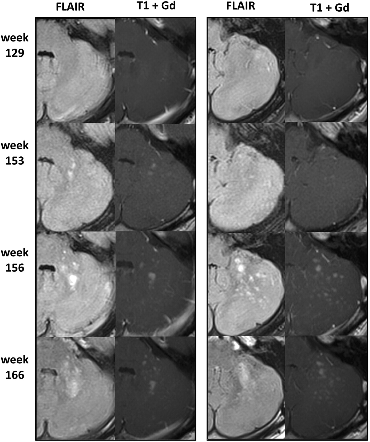
A 29-year-old man diagnosed 17 years earlier with relapsing multiple sclerosis (RMS) remained free of disease activity during 3 years of natalizumab therapy in that he experienced no clinical relapses, disability progression and no new T2 or gadolinium (Gd) enhancing T1 lesions on five successive follow-up brain MRI. He had no other significant comorbidities. The patient's previous RMS therapy included 4 years of interferon β-1b and six courses of mitoxantrone (12 mg/m2 dosed every 2 months, 3 years before initiating natalizumab), which failed to control disease activity. After 153 weeks of natalizumab therapy (38 doses of 300 mg intravenous every 4 weeks) our patient's sixth routine 6-month follow-up MRI scan depicted six small (1.0 to 3.0 mm) new lesions in the left middle cerebellar peduncle with faint T1 Gd enhancement (figure 1). Consequently, at the 39th scheduled natalizumab infusion (week 153), dosing was suspended, and a cranial MRI 3 weeks later demonstrated a major worsening of the punctuate-shape lesion pattern with 49 small (1.0–5.2 mm) Gd-enhancing lesions in the left middle cerebellar peduncle and left cerebellar hemisphere (week 156, figure 1). Most lesions were also hyperintense in diffusion-weighted imaging, and no oedema or meningeal enhancement was observed. The supratentorial MS lesions were strictly stable in number and volume with no Gd enhancement (figure 2, supplementary material). The only new complaint expressed by the patient was a mild occipital headache lasting 3–4 days, but his neurological examination remained unchanged and the Expanded Disability Status Scale score was stable at 4.0, as were several functional assessments such as the Timed 25-Foot Walk test,1 2 the Six-Spot-Step Test,3 the Timed 100 m Walk test,4 the Nine-Hole Peg Test1 2 and the 3 s Paced Auditory Serial Addition Test1 2 (figure 3, supplementary material). The cerebrospinal fluid analysis (CSF) showed a pleiocytosis with 81 white blood cells (91% lymphocytes, 9% monocytes) and eight red blood cells/mm3. The glucose concentration was within the normal range (0.64 g/l), and the protein concentration was mildly elevated (507 mg/l). The IgG was elevated at 50 mg/l, and numerous CSF-specific oligoclonal bands were observed. JC polyoma virus (JCV) DNA was detected in CSF using ultrasensitive quantitative real-time PCR (Focus Diagnostics, Cypress, California) (712 copies/ml), and serum anti-JCV antibodies were detected using a two-step ELISA as previously described.5 Other CSF and blood analysis were unremarkable (table 1, supplementary material). Based on these observations, we confirmed a diagnosis of asymptomatic progressive multifocal leucoencephalopathy (PML) in the posterior fossa, with an atypical punctuate-shape pattern (week 156). In order to hasten the clearance of natalizumab, we carried out only three plasma exchanges (PLEX, 1.5 volumes) since the last natalizumab infusion had been dosed, 7 weeks before PML diagnosis, with the possible presence of early MRI signs of immune reconstitution inflammatory syndrome (IRIS). Intravenous methylprednisolone (IVMP, 1 g) was initiated together with the PLEX for 5 days, followed by an oral tapering course of 12 weeks, in order to silence the radiological IRIS. After PLEX had been performed, we were informed that natalizumab serum concentration was already below the limits of quantitation of the assay (ie, <0.5 μg/ml) before PLEX. The patient remained strictly symptom-free throughout a follow-up of 19 weeks after the last natalizumab infusion, 15 weeks after the first MRI signs of PML and 12 weeks after PLEX (figure 2, supplementary material). Ten weeks after PML diagnosis and onset of PLEX-IVMP treatment, a follow-up assay using ultrasensitive PCR indicated that CSF was negative for JC virus DNA. The CSF pleiocytosis had vanished with four white blood cells/mm3, and protein concentration decreased at 426 mg/l. At the time the CSF was normalised (week 166), the follow-up brain MRI scans showed a regression of the PML lesion volume on T2/FLAIR sequences with a partial resolution of T1-Gd enhancement, particularly at the periphery of the lesion (figure 1). Brain MRI scans performed at weeks 157, 159 and 162 displayed intermediate lesion patterns consistent with the demonstration of a gradual radiological improvement between week 156 and 166 (data not shown).
T2-weighting with fluid attenuation inversion recovery (FLAIR) and T1+Gd MR images of two scanning plans separated by 8 mm in the posterior fossa at different stages of a patient's history with reference to the onset of natalizumab therapy. Week 129 examination was the fifth routine 6-monthly brain MR scan. Week 153 shows the first MR signs of the punctate-shape PML lesion in the left cerebellar peduncle. FLAIR-hyperintense dots (arrow) were all Gd-enhanced on T1-weighted images. At the time PML diagnosis was ascertained at week 156, the lesion massively extended to the left cerebellar hemisphere. When no more JC virus DNA was detectable on the CSF at week 166, we observed a significant regression of the multifocal lesion volume on FLAIR images together with an increased coalescence. The aspect of Gd enhancement was also modified and then restricted to the central core of the lesion with a decrease in dotted peripheral enhancement in the cerebellar hemisphere.
Discussion
Natalizumab is a highly effective therapy for RMS6 but increased exposure has been associated with an increased risk of PML, which is a potentially life-threatening central-nervous-system infection of oligodendrocytes by the JC polyoma virus.7 Despite an increased risk of PML in patients with known risk factors such as prior use of immunosuppressive agents and natalizumab treatment duration of more than 2 years, no clear guidelines have yet been drawn, and clinical vigilance remains the primary tool for early PML diagnosis. There is an ongoing debate about how to appropriately stratify PML risk in natalizumab-treated patients.5 8 It was recently reported that anti-JCV antibodies were detected in 100% of PML patients from whom pre-PML serum samples were available up to 187 months prior to clinical diagnosis,5 and as such, anti-JCV antibody serostatus is being investigated as potential additional factor to stratify PML risk.
Since the initial clinical manifestations of PML can be subtle (including isolated mild cognitive changes), it is not surprising that the time between the onset of symptoms and PML diagnosis can vary considerably, from a few days to several months.7 The management of natalizumab-associated PML is also a matter of debate. A prompt wash-out of serum natalizumab, either by PLEX or by immunoadsorption, in order to hasten the restoration of CNS immune surveillance, combined with steroid therapy to prevent IRIS development, is common practice for PML treatment. Recently, it has been reported in a group of 35 natalizumab-treated PML cases that enhanced clinical vigilance, including early recognition of symptoms, diagnosis and aggressive management of PML and IRIS are associated with an improved survival rate and clinical outcomes.9 However, the precise approach should be optimised for each patient with attention to MRI characteristics of PML (including early signs of IRIS) and of the natalizumab serum concentration at the time of PML diagnosis.
In our department, we perform 6-monthly brain MRI scans for all RMS patients treated with natalizumab. In the case of the patient reported here, the MRI features were somewhat unusual. In particular, the punctuate pattern is atypical and might be due to tiny multifocal foci of JC virus proliferation that later coalesce to give rise to the classical appearance of a widespread white-matter disorder and symptoms. Whereas the Gd enhancement is rare in HIV-related PML, it is commonly observed in the context of natalizumab therapy. Gd enhancement of the PML lesions may reflect a breakdown of the blood–brain barrier due to early IRIS10 associated with the rapid decline of natalizumab serum concentration, which was already undetectable here when the first lesions were observed. Finally, the rapid deterioration of the PML lesion load observed within 3 weeks after the first MRI signs is a strong argument to plead for a rapid CSF screening for JC virus DNA in the context of any new unusual brain lesion in MS patients treated with natalizumab.8
This case emphasises that JC virus CNS infection can be detected at a preclinical stage, even in the posterior fossa where small lesions often cause serious disability. The uncoupling between a large PML lesion in a highly functional location, and the absence of clinical expression, is of great interest. In this case, an aggressive corticosteroid regimen initiated during PLEX was associated with a stable clinical course of PML, a favourable evolution of MRI manifestations and clearance of JC virus from CSF. Although the benefits of steroids in natalizumab-associated PML-IRIS have never been and will never be addressed in a randomised clinical trial, at least one study has suggested a clinical benefit in PML-IRIS with HIV infection.11 Since the theoretical risk of increasing the JC virus load during steroid therapy is considered low with regard to the likelihood of developing detrimental IRIS, many experts have advocated high-dose steroid treatment use in the context of natalizumab-associated PML.7 12 13
One prior reported case suggests that asymptomatic MRI changes can be captured several months prior to initial manifestation of PML symptoms.14 The story of the present case suggests that the screening for preclinical signs of PML might be achieved through periodic T2/FLAIR brain MRI scans in patients with known PML risk factors such as prior immunosuppressant exposure, natalizumab treatment duration over 2 years and JC virus seropositivity. However, the required frequency, cost–benefit trade-off and efficacy of such a monitoring remain to be investigated before we firmly consider that it may increase the proportion of early detection of PML in such patients, and improve their chance of avoiding or minimising permanent neurological injury.
References
Supplementary materials
Supplementary Data
This web only file has been produced by the BMJ Publishing Group from an electronic file supplied by the author(s) and has not been edited for content.
Files in this Data Supplement:
- Download Supplementary Data (PDF) - Manuscript file of format pdf
Supplementary Data
This web only file has been produced by the BMJ Publishing Group from an electronic file supplied by the author(s) and has not been edited for content.
Files in this Data Supplement:
- Download Supplementary Data (PDF) - Manuscript file of format pdf
Footnotes
Competing interests None.
Patient consent Obtained.
Ethics approval Ethics approval was provided by the ethical committee of the CHU of Liège, Belgium.
Provenance and peer review Not commissioned; externally peer reviewed.