Article Text

Abstract
Lacunar stroke is a marker of cerebral small vessel disease and accounts for up to 25% of ischaemic stroke. In this narrative review, we provide an overview of potential lacunar stroke mechanisms and discuss therapeutic implications based on the underlying mechanism. For this paper, we reviewed the literature from important studies (randomised trials, exploratory comparative studies and case series) on lacunar stroke patients with a focus on more recent studies highlighting mechanisms and stroke prevention strategies in patients with lacunar stroke. These studies suggest that lacunar stroke is a heterogeneous disease with various mechanisms, including most commonly lipohyalinosis and less commonly atheromatous disease and cardioembolism, highlighting the importance of a careful review of brain and neurovascular imaging, a cardiac and systemic evaluation. A better understanding of pathomechanisms of neurological deterioration may lead to investigating the utility of novel treatment strategies and optimisation of short-term antithrombotic treatment strategies to reduce the risk of neurological deterioration and prevent long-term disability in patients with lacunar stroke.
- stroke
Statistics from Altmetric.com
Introduction
Lacunar stroke is a marker of cerebral small vessel disease1 and accounts for up to 25% of ischaemic stroke. The word lacunar comes from Latin for ‘lacuna’ meaning hole, and it is used to describe a small focus of encephalomalacia containing CSF, which is the end result of liquefactive necrosis. Lacunar stroke is defined as a subcortical infarct measuring less than 20 mm in diameter, caused by occlusion of a perforator of an intracranial artery.1 In this narrative review, we aim to provide an overview of potential lacunar stroke mechanisms and diagnostic approaches, and discuss therapeutic implications targeting the underlying mechanism.
Background
The incidence of lacunar stroke varies based on the population studied from 25 to 50 per 100 000 people,2 3 comprising 15%–25% of ischaemic stroke.2–4 These numbers, however, have been declining over time, likely due to better control of vascular risk factors such as hypertension.5
Lacunar stroke shares risk factors with other stroke subtypes, namely hypertension, diabetes, advanced age, cigarette smoking and hyperlipidaemia.6 7 While studies have shown that the overall prevalence of these risk factors is similar between lacunar stroke and other stroke subtypes,8 some studies suggest that smoking, hypertension and diabetes are particularly important risk factors for lacunar stroke3 7 and that these risk factors may be more prevalent in patients with lacunar stroke.9 Among these risk factors, hypertension is most common in patients with lacunar stroke (68%), followed by diabetes (30%).3 7 9 These studies were performed when the control of risk factors, particularly hypertension, was less aggressive and more recent studies suggest that risk factors for lacunar stroke may be similar to those of other subtypes.10
In addition to conventional risk factors, rare genetic conditions, such as Cerebral Autosomal Arteriopathy with Subcortical Infarcts and Leukoencephalopathy (CADASIL) can cause lacunar stroke.11 These typically have other accompanying manifestations, including a positive family history, and the diagnosis is made by clinical suspicion and confirmed by genetic testing (table 1).11
Overview of genetic conditions associated with lacunar stroke
Potential mechanisms
There are several potential mechanisms described for the pathogenesis of lacunar stroke.12 These include lipohyalinosis, atherosclerotic disease and cardiac embolism, which will be discussed separately below.
Lipohyalinosis
Lipohyalinosis is defined as concentric hyaline thickening of the cerebral small vessels leading to occlusion of the small penetrating arteries13 and is one of the first and most common lacunar stroke mechanisms described and pathologically proven to cause lacunar stroke.14 Lipohyalinosis is thought to originate from hypertension-related hypertrophy and fibrinoid degeneration of the vessel walls as well as subintimal foam cells obliterating the lumen of small penetrating arteries, leading to small subcortical infarcts. In a case series published by Fisher of 114 lacunar strokes, all but three had direct or indirect evidence of uncontrolled hypertension.14 Previous studies suggested that lipohyalinosis typically causes infarcts 3–7 mm in size on brain imaging and that if the infarct is larger than 7 mm, other mechanisms should be explored.15
In addition to lipohyalinosis, some studies hypothesised that endothelial dysfunction and impaired autoregulation as well as extravasation of blood products into the vessel wall resulting in perivascular oedema and damage to the neurovascular unit and surrounding brain tissue may be a contributing factor to the development of lacunar infarcts.13 This is hypothesised to lead to lacunar stroke and white matter disease.16 This mechanism has not been pathologically proven and recently data suggests that endothelial dysfunction may be reactive and less likely to be involved in the pathogenesis of the disease itself.17
Atherosclerotic disease
There are several potential atherosclerotic mechanisms that could lead to lacunar stroke. The most important mechanism that has been pathologically proven is branch atheromatous disease.18 Atherosclerotic plaques of the parent artery could involve the ostium of perforating branches leading to occlusion and infarction of distal parenchyma. This mechanism has been widely described in patients with luminal narrowing of the parent artery, in which case the lacunar stroke is classified as related to intracranial atherosclerosis as opposed to small vessel disease. On the other hand, certain atherosclerotic plaques may cause perforator disease without significant luminal stenosis. This has been described particularly in patients with pontine lacunar stroke in the setting of branch atheromatous disease of the basilar artery.19
Other atherosclerotic mechanisms that have been described include embolism from a proximal intracranial or extracranial artery20 as well as aortic arch disease.21 These associations, however, do not prove a causal relationship with lacunar stroke because atherosclerosis and lacunar stroke share common risk factors.
Cardioembolism
There is experimental evidence from animal models that small emboli can enter penetrating arteries and cause lacunar stroke.22 In humans, there is evidence to suggest that cardioembolic mechanisms are unlikely to cause small subcortical infarcts. For instance, one study showed that atrial fibrillation (AF) patients with lacunar stroke are more likely to have increased white matter disease severity and evidence of chronic lacunar stroke than those with cardioembolic-appearing infarcts, suggesting that even in the presence of AF, intrinsic lipohyalinosis is a more likely aetiology.23 Furthermore, studies have shown that subcortical infarcts are less likely to occur in the setting of a patent foramen ovale.24 Therefore, current evidence suggests that lacunar stroke is a very rare manifestation of cardioembolism.
Diagnostic approach
Brain imaging and non-invasive intracranial vascular imaging
Infarct location on brain imaging could help determine the mechanism in lacunar stroke. For instance, one study showed that a lacunar stroke involving the paramedian thalamus could be related to a distant cardioaortic embolic source25 but more studies are needed to confirm this finding. Obtaining a brain MRI may be particularly helpful not only to confirm the diagnosis but also to help determine the aetiology. For instance, the presence of deep cerebral microbleeds, prior subcortical lacunar infarcts or subcortical white matter disease are better appreciated on brain MRI and may suggest an intrinsic small vessel microangiopathy as opposed to a distant atheroembolic or cardioaortic source. On the other hand, acute infarcts in more than one vascular territory may suggest a proximal cardioarotic source. Future studies are needed to determine the cost-effectiveness of obtaining routine brain MRI versus CT in patients with lacunar stroke.
Furthermore, preliminary data suggests that increased pulsatility index, a marker of arterial stiffness, in the basal ganglia on 7T MRI was more common in patients with lacunar stroke and deep intracerebral haemorrhage than controls indicating the need for further studies to determine whether increased pulsatility index could unravel the underlying mechanism in patients with lacunar stroke.26 A major limitation of this approach is that 7T MRI is not used in routine clinical care and thus the clinical utility of this remains limited.
Vessel imaging
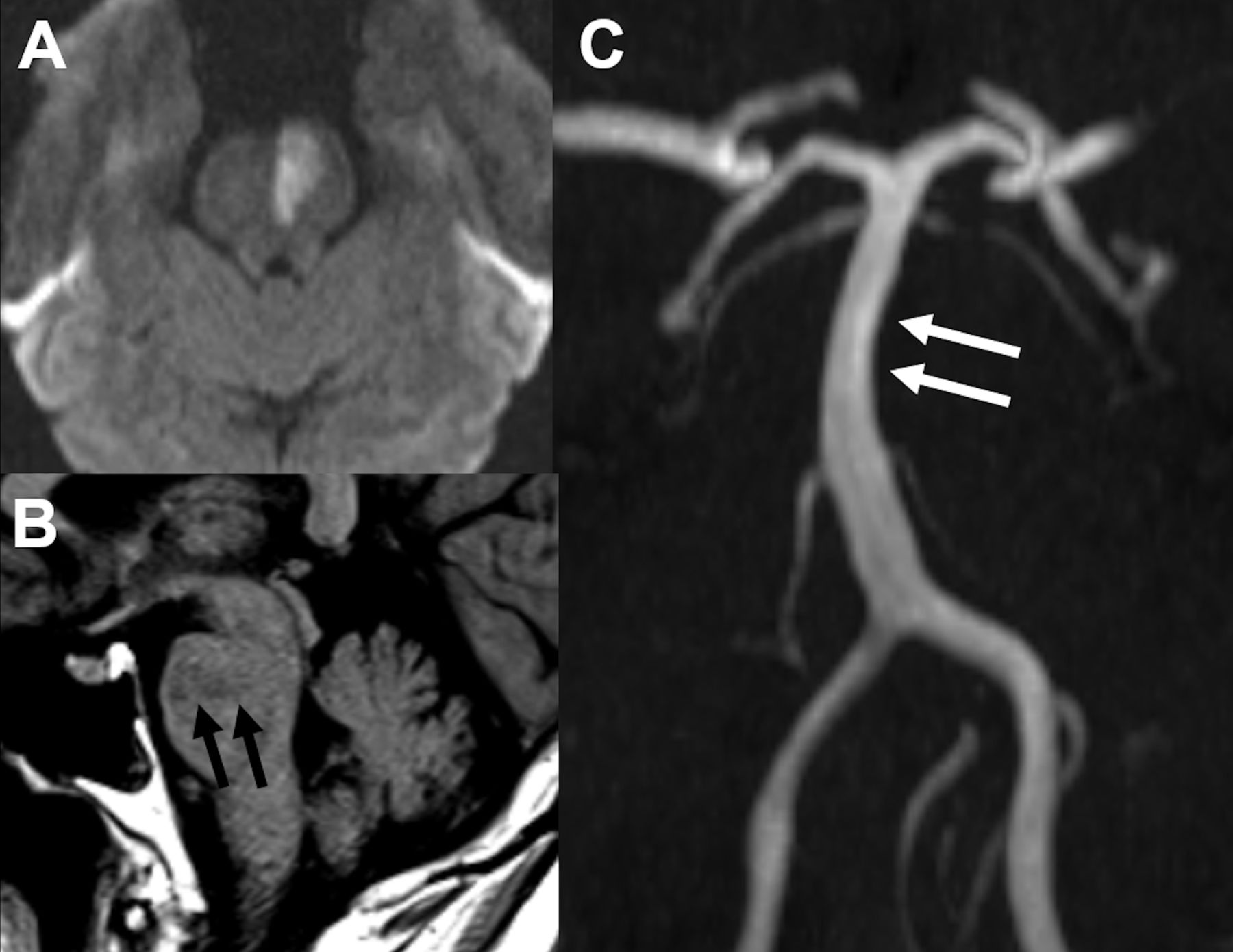
Non-invasive intracranial vessel imaging may also help determine the aetiology of a lacunar infarct. For instance, irregularity or stenosis on intracranial MRA or CTA in the main artery at the site where the perforating branch supplying the infarct originates may suggest an underlying atherosclerotic mechanism and ischaemic lesion patterns may differ between atherosclerotic and non-atherosclerotic mechanisms (figure 1).27 In one study, 40% of patients with small striatocapsular infarcts were found to have stenosis of the middle cerebral artery.28

Brainstem lacunar infarct from basilar atherosclerosis. diffusion weighted imaging sequence (A), sagittal T1 (B) and MPR from TOF MRA (C). Notice the acute infarct involving the typical territory (black arrows on B) of a paramedian basilar perforator which arise from the mid basilar artery; atherosclerosis of the basilar artery with mild stenosis is well evident on the MRA, asymmetrical to the left side (white arrows on C). MPR, multiplanar reconstruction; MRA, Magnetic Resonance Angiography
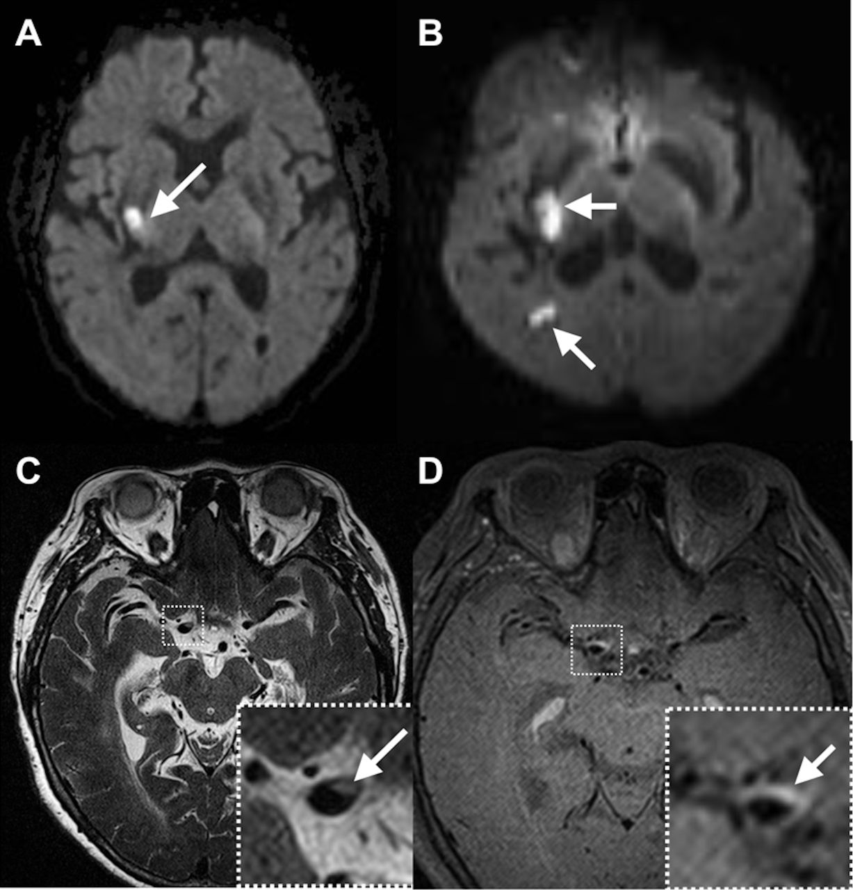
Vessel wall imaging (VWI) is a promising MRI sequence that can be added to the standard MRI of the brain and uses signal suppression from the tissues surrounding the intracranial arterial wall, namely the blood and CSF. Atherosclerotic plaques typically are associated with eccentric non-uniform arterial wall thickening, usually T2 hyperintense and enhancing in the setting of recent stroke.29 VWI may help with the differentiation between the two aetiologies described above—lipohyalinosis and perforator origin atherosclerosis (figure 2).30 Other VWI studies have focused on analysis of the plaque location compared with the lacunar stroke, confirming that MCA atherosclerotic plaques with a lacunar stroke have more involvement of the superior wall,31 but this finding needs confirmation in larger studies.

Middle-aged patient with history of hypertension and hyperlipidaemia presented with left arm weakness and facial droop. MRI in panel A revealed a small lacunar appearing stroke (white arrow) in the right internal capsule. Two days later, she developed more severe left-sided weakness and a repeat MRI in panel B showed extension of the prior stroke and a second stroke in a more distal location in the right parietal lobe. The second stroke instigated a thorough workup, including CTA of the head and neck, transthoracic echocardiogram with bubble study, hypercoagulable panel and extended rhythm monitoring, which all failed to reveal the cause of the patient’s strokes. A vessel wall MRI revealed an inflamed atherosclerotic plaque adjacent to the origin of the lenticulostriate perforators responsible for the initial stroke, shown in C on T2-weighted SPACE with DANTE black blood prepulacunar strokee, which enhanced avidly after gadolinium administration, shown in D on T1-weighted SPACE with DANTE black blood prepulacunar strokee. This plaque was determined to be the cause of the patient’s strokes and after being placed on aggressive medical management with DAPT, there was no stroke recurrence. DAPT, dual antiplatelet therapy.
Currently, using vascular imaging and VWI to determine an atherosclerotic mechanism in patients with lacunar stroke may not necessarily lead to a change in secondary stroke prevention strategies. This, however, may change if future studies show a benefit from targeted treatment strategies such as anti-inflammatory agents as well as novel lipid-lowering agents for secondary stroke prevention in patients with cerebrovascular atherosclerosis.
Cardiac evaluation
The utility of cardiac evaluation in patients with lacunar stroke is a subject of interest. This includes echocardiography and outpatient cardiac monitoring to evaluate for paroxysmal occult AF. A recent study challenged the yield of transthoracic echocardiography (TTE) in patients with lacunar stroke.32 One study showed that an aortic arch atheroma detected on transesophageal echocardiographic (TEE) is more likely to occur in patients with lacunar stroke compared with controls but this finding has not been validated. In addition, detecting an aortic atheroma rarely leads to a change in clinical management because stroke prevention strategies are essentially similar in patients with small vessel disease and complex aortic arch atheromas.21
In addition, a recent meta-analysis showed that AF detection, with at least 7 days of cardiac monitoring, was significantly higher in patients with cryptogenic stroke versus lacunar stroke subtype (9.2% vs 2.4%, p=0.02).33 The 2.4% AF detection rate in patients with lacunar stroke in this study may not be trivial and requires more study. The ‘Rate of Atrial Fibrillation Through 12 Months in Patients With Recent Ischaemic Stroke of Presumed Known Origin’ (Stroke-AF) trial showed increased AF detection at 12 months with insertable cardiac monitor versus standard of care in patients with lacunar stroke (HR 13.83 95% CI 1.80 to 111.11, p<0.001) with a 12.6% AF detection at 1 year (NCT02700945). It remains unclear, however, whether anticoagulation therapy in those who were found to have brief subclinical AF has a secondary prevention benefit.
In conclusion, although the yield of echocardiography and cardiac monitoring in patients with lacunar stroke is low, some patients with a suspected distant embolic source based on neuroimaging (such as lack of other signs of small vessel disease) or younger patients without vascular risk factors may benefit from a cardiac diagnostic evaluation including TTE and possibly TEE as well as outpatient cardiac rhythm monitoring in certain patients to determine a potential cardiac mechanism. Ongoing trials will shed light on the utility of extended cardiac monitoring in patients with lacunar stroke.
Genetic testing
While not common, certain genetic conditions such as Cerebral autosomal dominant arteriopathy with subcortical infarcts and leukoencephalopathy (CADASIL), Cerebral autosomal recessive arteriopathy with subcortical infarcts and leukoencephalopathy (CARASIL) and Collagen Type IV Alpha 2 Chain (COL4A2) may cause lacunar stroke. These conditions should be considered in patients with a lacunar infarct with minimal or no risk factors and the presence of other manifestations of these conditions as well as a positive family history, particularly if there is evidence of other markers of intrinsic small vessel disease.34 In addition, a thoughtful approach that weighs the cost versus benefit of diagnosing such conditions particularly if treatment is available or for prognosis and family planning purposes, is crucial before these tests are ordered. Furthermore, a recent international pooled analysis of patients with MRI confirmed lacunar stroke identified 12 genetic loci that may be implicated in the pathogenesis of lacunar stroke and may represent future therapeutic targets.35
Looking beyond the brain
A comprehensive diagnostic evaluation is important to look for evidence of small vessel disease in other organs such as the kidneys, heart, and retina. For instance, one study showed that patients with cerebral small vessel disease were more likely to have changes in the retinal vasculature, suggesting that pathologic changes in the retinal arteries parallel changes in the small cerebral arteries, even in normotensive patients.36 In addition, recent evidence suggest that small vessel disease is a multisystem disorder that may the culprit in ‘cardiac syndrome X’ or ‘microangina’ as well as chronic kidney disease and ischaemic nephropathy.37 This concept is further supported by genetic cerebral small vessel disease disorders such as CADASIL which also affects other organs such as the skin and heart.37 These data underscore the importance of a comprehensive multisystem evaluation in patients with lacunar stroke which may not only help determine the likely stroke mechanism but also lead to changes in management of other organs such as referral to an ophthalmologist in patients with evidence of retinopathy.
Neurological deterioration
Studies suggested that 23%–41% of patients with lacunar stroke exhibit neurological deterioration after symptom onset.38–40 The definition of neurological deterioration and the inclusion/exclusion criteria varied across these studies thus the rate may be on the higher end with less conservative definitions and lower end with more conservative definitions. Studies have shown that patients with lacunar stroke who exhibit neurological deterioration have increased odds of poor functional outcome at follow-up.38–41 Identifying potential mechanisms for neurological deterioration may guide strategies to prevent and treat neurological deterioration in lacunar stroke.
Plausible mechanisms
Mechanisms hypothesised to be in the pathogenesis of neurological deterioration after lacunar stroke include branch atheromatous disease, altered haemodynamics, disruption of neurovascular unit resulting in oedema, excitotoxicity and inflammation.
There are data to suggest that branch atheromatous disease is associated with increased risk of neurological deterioration in patients with lacunar stroke. In several studies, the odds of early neurological deterioration in patients with small subcortical infarcts was increased with the presence of branch atheromatous disease.42 On the other hand, markers of small vessel disease such as cerebral microbleeds and white matter disease severity were not associated with the risk of neurological deterioration.43 The proposed mechanism is that the atherosclerotic plaque extends to involve more penetrating arteries causing the infarct to expand. As such, patients with branch atheromatous disease have larger infarcts compared with those without branch atheromatous disease.44
Haemodynamic failure is another proposed mechanism of neurological deterioration in patients with lacunar stroke. In one study, patients with lacunar stroke who exhibited neurological deterioration were found to have lower cerebral blood flow and increased mean transit time in the region of interest.45 At least 20% of patients with lacunar stroke have an ischaemic penumbra on perfusion weighted imaging,46 47 which is also associated with neurological deterioration.47 It remains unclear however whether this association is related to the perfusion abnormality or whether larger initial infarcts are associated with more readily detected penumbra and with greater likelihood of deterioration. The theory of impaired perfusion is further supported by evidence showing that impaired cerebrovascular reactivity is more common in lacunar stroke patients who exhibit neurological deterioration.48
Other mechanisms hypothesised to be implicated in the pathogenesis in neurological deterioration are oedema, excitotoxicity, and inflammation. It has been hypothesised that oedema caused by the disruption of the blood brain barrier can lead to compression of small perforators near the ischaemic lesion causing infarct extension and neurological deterioration.49 Another study showed increased serum concentration of glutamate and decreased serum concentration of Gamma aminobutyric acid in patients with lacunar stroke who exhibited neurological deterioration, suggesting that excitotoxicity may be another mechanism of neurological deterioration.50 Neurological deterioration may also occur in the setting of systemic inflammation and thrombosis. This is supported by studies showing higher levels of inflammatory biomarkers41 51 and cytokines52 in those who exhibit neurological deterioration.
Therefore, the exact pathogenesis of neurological deterioration in patients with lacunar stroke remains poorly understood highlighting the need of large observational studies using advanced imaging modalities such as perfusion, VWI, cerebrovascular reactivity in addition to measurement of serum markers of excitotoxicity and inflammation to better understand these mechanisms. A better understanding of the pathogenesis of neurological deterioration may lead to investigating therapeutic strategies to treat and prevent neurological deterioration.
Secondary prevention
Pharmacological interventions
Pharmacological interventions53–61 including use of antithrombotic agents, blood pressure (BP) lowering agents, statin therapy, glycaemic agents, as well treatment of insulin resistance for secondary prevention in patients with lacunar stroke are summarised in table 2. Interest is given to the promising role of cilastozol which is a phosphodiesterase III inhibitor that induces mild antiplatelet effects, BP reduction and reduction in triglycerides for stroke prevention after lacunar stroke.
Summary of pharmacological secondary prevention strategies in patients with lacunar stroke
Lifestyle
Key to long-term prevention of recurrent lacunar stroke is sustained control of risk factors, namely hypertension, physical inactivity, tobacco use, diabetes and hyperlipidaemia. Physical activity plays an important supporting role in secondary stroke prevention, as increased physical activity decreases BP, promotes lower HbA1C (when part of a structured programme), reduces insulin resistance, improves the lipid profile and may reduce stroke risk itself, among other positive effects on chronic disease.62 Current recommendations for adults are 150 min of moderate intensity aerobic physical activity weekly.63 Because low rates of exercise participation are common in stroke survivors, targeted inquiries into reasons for decreased activity and patient-specific recommendations may better address this missed opportunity. Furthermore, smoking is a risk factor for lacunar stroke and thus smoking cessation is an important intervention in reducing the risk of recurrent stroke and cardiovascular events.63 64
Future directions: preventing neurological deterioration and targeting the underlying mechanism
Prevention and treatment of neurological deterioration
For patients with stuttering lacunar stroke, there may be a role for early dual antiplatelet therapy (DAPT) with aspirin and clopidogrel.65 In a recently published retrospective study of 458 patients with lacunar stroke, 130 (28%) of patients had stuttering symptoms that met the definition of early neurologic deterioration. DAPT treatment reduced stuttering symptoms from 77% of patients receiving antiplatelet monotherapy to only 21% of patients receiving DAPT.40 Specifically in patients with lacunar stroke and evidence of branch atheromatous disease, a multicentre observational study showed that when compared with aspirin monotherapy, aspirin plus cilostizol within 12 hours from onset was associated lower likelihood of clinical progression.66 This study, however, is open label and non-randomised and results should be interpreted with caution given the potential for bias. The LACunar Intervention Trial-1 (LACI-1) trial of 57 patients with lacunar stroke in the UK showed that the combination of cilostazol and isosorbide mononitrate was safe and well tolerated.67 The ongoing LACI-2 is testing the feasibility of conducting a large phase 3 trial to investigate the safety and efficacy of cilostazol and/or isosorbide mononitrate in patients with lacunar stroke.68 Other small studies have looked at the use of intravenous glycoprotein IIb/IIIa inhibitors to prevent deterioration in stuttering lacunar stroke, with promising preliminary results.69 70 Nonetheless, additional data is needed to support their use, considering that DAPT with a loading dose has a potential benefit and lower cost.
Two retrospective studies in Korea reported a benefit for induced hypertension with phenylephrine in patients with stuttering lacunar stroke.71 72 The sample size in these studies was small and conclusions cannot be drawn apart from the preliminary safety of mild induced hypertension. Nonetheless, there is biological plausibility to the concept that inducing hypertension could be beneficial in stuttering lacunar stroke, as it may maximise perfusion of the penumbral region. In the IMAGES study, which randomised 2589 patients to either an intravenous magnesium or placebo within 12 hours of stroke onset, a subgroup analysis of patients with lacunar stroke showed that although the overall IMAGES study was neutral, in 765 patients with lacunar stroke there was benefit for magnesium.73 The OR for favourable outcome (modified Rankin Scale 0–1 at 90 days) in lacunar stroke patients who received magnesium was 0.70 (95% CI 0.53 to 0.92) and the interaction term between lacunar stroke and magnesium therapy was significant (p=0.005). While subgroup analyses of a clinical trial should be interpreted with caution, the results may warrant additional study given the potentially neuroprotective effects of magnesium on cerebral white matter.74
Targeted treatments for lacunar stroke in the setting of substenotic atherosclerosis
Studies have shown that in patients with cardiovascular atherosclerotic disease, optimising antithrombotic therapy with the use of low dose rivaroxaban was associated with a reduced risk of major cardiovascular events.75 The benefit of this treatment in patients with symptomatic stenosing intracranial atherosclerosis or non-stenosing branch atheromatous disease remains uncertain. This was also the case with anti-inflammatory drugs such as low dose colchicine76 and canakinumab77 and lipid lowering agents such as proprotein convertase subtilisin/kexin type 9-inhibitors.78 In addition, the ‘Treat Stroke to Target’ trial showed that in patients with an ischaemic stroke or TIA and evidence of atherosclerosis, a target low-density lipoprotein cholesterol <70 mg/dL had a lower risk of subsequent cardiovascular events compared toa target range of 90–110 mg/dL.79 These could be investigated in future trials aiming to reduce the risk of recurrence in patients with lacunar stroke whose mechanism is thought to be related to substenotic atherosclerosis.
Lacunar stroke and risk factors for cardioembolism
In patients whose lacunar stroke occurs in the setting of known paroxysmal or permanent AF, anticoagulation is recommended for secondary stroke prevention. Several randomised trials have shown that DOACs are non-inferior to warfarin at prevention of thrombotic events, with lower risk of intracranial haemorrhage.80 This may be particularly important in patients with lacunar stroke and concomitant microbleeds whose risk of intracerebral haemorrhage may be particularly increased. In such patients with biomarkers of haemorrhagic risk such as high microbleed burden or severe white matter disease, studies investigating the safety and efficacy of left atrial appendage occlusion compared with DOACs are needed.
While lacunar strokes are unlikely to be related to a distant embolic source, in young patients who have a lacunar stroke and a PFO in the absence of AF and risk factors for cerebral small vessel disease,81 PFO closure may be reasonable (figure 3).

Diffusion-weighted imaging sequence showing a lacunar infarct in a young patient without risk factors who presented with acute onset right sided weakness. Diagnostic evaluation including telemtery, vascular imaging, hypercoagylable labs and echocardiography revelled a patent formen ovale (PFO) with a bidirectional shunt. His Risk of Paradoxical Embolism (ROPE) score was nine suggesting that the PFO is the likely culprit.
Conclusion
Lacunar stroke is a common and heterogeneous disease that necessitates a nuanced approach to diagnosis and treatment. Control of secondary stroke risk factors, principle among them hypertension, is a critical element in lowering the risk of recurrent lacunar stroke. Primary prevention of lacunar stroke has not been studied, but ongoing studies in patients with asymptomatic brain lesions will provide much needed data regarding the safety and efficacy of aggressive medical management to prevent stroke, of which lacunar stroke is the main symptomatic subtype.
Ethics statements
References
Footnotes
Twitter @eytanraz
Contributors SY: manuscript preparation, revision, concept and design ER and AdH: manuscript preparation, revision and imaging acquisition. DY, BMG and SC: manuscript preparation and revision. MSVE: manuscript revision.
Funding The authors have not declared a specific grant for this research from any funding agency in the public, commercial or not-for-profit sectors.
Competing interests None declared.
Provenance and peer review Not commissioned; externally peer reviewed.