Article Text

Abstract
Objectives: The evolutionary pattern of spontaneous recovery from acute neglect was studied by assessing cognitive deficits and motor impairments. Detailed lesion reconstruction was also performed to correlate the presence of and recovery from neglect to neural substrates.
Methods: A consecutive series of right brain-damaged (RBD) patients with and without neglect underwent weekly tests in the acute phase of the illness. The battery assessed neglect deficits, neglect-related deficits, and motor impairment. Age-matched normal subjects were also investigated to ascertain the presence of non lateralised attentional deficits. Some neglect patients were also available for later investigation during the chronic phase of their illness.
Results: Partial recovery of neglect deficits was observed at the end of the acute period and during the chronic phase. Spatial attention was impaired in acute neglect patients, while non spatial attentional deficits were present in RBD patients with and without acute neglect. A strong association was found between acute neglect and fronto-parietal lesions. Similar lesions were associated with neglect persistence. In the chronic stage, neglect recovery was paralleled by improved motor control of the contralesional upper limb, thus emphasising that neglect is a negative prognostic factor in motor functional recovery.
Conclusions: These findings show that spatial attention deficits partially improve during the acute phase of the disease in less than half the patients investigated. There was an improvement in left visuospatial neglect at a later, chronic stage of the disease, but this recovery was not complete.
- MA, maximal amplitude
- RBD, right brain-damaged
- RT, reaction time
- attention
- lesion
- neglect recovery
- parietal
Statistics from Altmetric.com
Neglect patients most frequently fail in the acute phase to respond to contralesional stimuli following right hemisphere damage,1–3 although many patients may ignore contralesional stimuli for months or even years after the lesion.4–8 Despite the theoretical relevance of neglect evolution and its practical implications for rehabilitation, little is known about the pattern of spontaneous recovery of neglect. This study addressed four main questions. First, which attentional component (for example, spatial, sustained) is specifically impaired in acute neglect? Second, which aspects of the syndrome recover spontaneously in the acute phase (<6 weeks after stroke)? Third, how many patients recover in the acute and more chronic phases (>3 months after stroke), and which characteristics differentiate these patients from those who do not recover? Fourth, what are the anatomical correlates of acute and persistent neglect?
Regarding the first issue, both spatial and non spatial deficits were investigated.9–12 If disorders of sustained and divided attention play a specific role in determining neglect, then right brain-damaged (RBD) patients with neglect should be more impaired than patients without neglect in tests probing these attentional components.
Second, the spontaneous recovery patterns of spatial and non spatial attention were studied for a 2 week period during the acute phase because, if they reflect independent impairments, they may show different recovery patterns. A large battery of tests was used, as neglect may affect the personal or extrapersonal (near-far) space13–16 and the “perceptual” or “premotor” component,17–22 and may be associated with anosognosia, extinction, and primary sensory-motor deficits.23 Indeed the estimated frequency of neglect varies considerably with the type of test or coding criteria.24–29 Computerised tasks were used to assess perceptual-premotor components of neglect30–34 and the presence, severity, and evolution of anosognosia35,36 and extinction37–41 were also evaluated. If anosognosia and extinction are independent of neglect, their presence and recovery patterns might dissociate, otherwise they should coexist and recover similarly. Finally, as neglect may largely prevent motor recovery and functional independence,42,43 the severity and evolution of motor disabilities were assessed to verify whether motor improvement is associated or not with neglect recovery.
Third, we evaluated the proportion and the neuropsychological characteristics of acute patients who recovered significantly compared to those who did not recover during the same period. To this aim, 33 acute RBD patients (23 with and 10 without neglect) were examined three times over 2 successive weeks. A subgroup of eight patients was available for a chronic follow up examination.
Finally, the anatomical correlates of acute and persistent (no-recovery) visuospatial neglect were investigated. Neglect is often found after right temporo-parietal-occipital lesions.44–46 While parietal damage is the most frequent observation,2 neglect is also reported after lesion of the frontal lobe47 and superior/middle temporal gyrus.3,48 The role played by the superior temporal gyrus has been recently challenged by Mort and colleagues49 who, in a detailed study of the visual neglect anatomy, identified the angular gyrus in the inferior parietal lobe as the area critically associated with neglect. Neglect can also emerge after subcortical lesions,50–52 although it is possibly determined by concurrent cortical hypoperfusion.53–57 Some studies failed to relate the size of parietal lobe damage to neglect severity58–60; this relationship may be stronger during the acute than post-acute stage, although it appears difficult to distinguish between transient and persisting neglect on the basis of lesion location.61 Here we tried to clarify the anatomo-functional relationships between lesion location and the presence/absence of neglect and neglect recovery. To this aim, the method introduced by Damasio and Damasio62 was used for 29 out of the 33 RBD patients investigated (21 with and eight without neglect; CT/MRI scans of four patients were not available).
METHODS
A total of 33 acute patients (<6 weeks from stroke), selected from a larger group,63 were enrolled in the study after giving informed consent, provided that they had not suffered from previous neurological or DSM-IV Axis I disorders. Arousal and behavioural control were adequate for a 60 min testing session which included tests of spatial and non spatial attention, neglect-related disorders, and motor abilities. The order of the tests was randomised for each patient, and maintained throughout sessions. A left/right differential score (⩾20%) on accuracy in (bell or letter) cancellation tasks determined the presence of neglect, identifying (table 1) a neglect group of 23 patients (11 male, 12 female; mean age: 68 years; mean education: 11 years) and a no-neglect group of 10 patients (five male, five female: mean age: 72 years; mean education: 7 years). For comparative purposes concerning non spatial attentional deficits, 10 healthy subjects (five male, five female; mean age: 68 years; mean education: 6 years) were tested. Neglect group education was higher than that of the no-neglect and healthy groups (both p<0.04), the mean age being comparable.
Details of acute right brain-damaged patients
Material
Non spatial attention
In the Sustained Attention to Response Test (SART64) patients performed speeded key-press responses to each centrally presented digit (1–9) but one (3). Accuracy (go, no-go) and reaction time (RT, within 150–10 000 ms) were recorded. Arousal and divided attention were assessed with a simple RT task performed with and with out digit load (base-dual task65). Speeded key-press responses were required upon detection of a dot (randomly displayed on a computer screen), the dual task requiring concurrent repeat of a list of heard digits (whose span was pre-determined for each patient). Correct RTs (within 150–10 000 ms) were recorded.
Spatial attention
Four sub-tests of the BIT battery66 (letter cancellation, line bisection, picture scanning, and menu reading) as well as the bell cancellation task67 and fluff test (personal neglect68) were used. Accuracy scores were derived from each test. Deviations (in mm) to the right (+) or the left (−) of the objective midpoint were measured for the line bisection task.
Perceptual-premotor components of neglect were assessed by computerised tests.69 The lateralised target task asked the patient to make a speeded key-press response (space bar) when a downward oriented arrow randomly replaced one of three dots initially displayed (left, centre, right), irrespective of its position on the screen. In the lateralised response task only the central dot was replaced by the arrow (randomly oriented to the left, right, or downward), and patients were required to press as fast as possible a left, a right, or a central button, according to the orientation of the arrow. RTs and omissions were recorded.
Neglect-related disorders
Patients’ awareness of sensorimotor impairments was assessed by a questionnaire70 containing typical questions such as: Is there anything wrong with your arm? your leg? A score from 0 to 10 (absent to severe anosognosia) reflected deviation of actual from reported impairment. Visual and tactile extinction was assessed by confrontation.
Motor abilities
Joint range of motion was assessed (in degrees) for contralesional arm movements including: shoulder flexion (maximal amplitude (MA) 180°), shoulder abduction (MA 180°), elbow flexion (MA 80°), wrist extension (dorsi-flexion, MA 90°), and finger metacarpo-phalangeal extension (MA 90°). The contralesional grip force was measured with a dynamometer (Lafayette Instruments).
Procedure
Patients were assessed three times, at weekly intervals: at recruiting, that is, 1–6 weeks after stroke (first session; table 1), and then 1 and 2 weeks after (second and third sessions). A subgroup of eight neglect patients underwent a fourth follow-up session (without computerised tests) in the chronic phase (>3 months after stroke). Age-matched normal subjects performed the SART and base-dual tests in a single session.
Anatomical correlates of acute neglect
Lesions were plotted against brain templates from the atlas of Damasio and Damasio,62 identifying 45 anatomical structures with respect to which CT/MRI scans were judged. Lesions were first manually transcribed onto templates (A18 or A20), singly selected according to the appropriate scan plane (figs 1–3). Then, digitised images of reconstructed lesions of all neglect (fig 1) and no-neglect (fig 2) patients were separately plotted onto the same templates, showing the area of maximum overlap on each slice of each template.
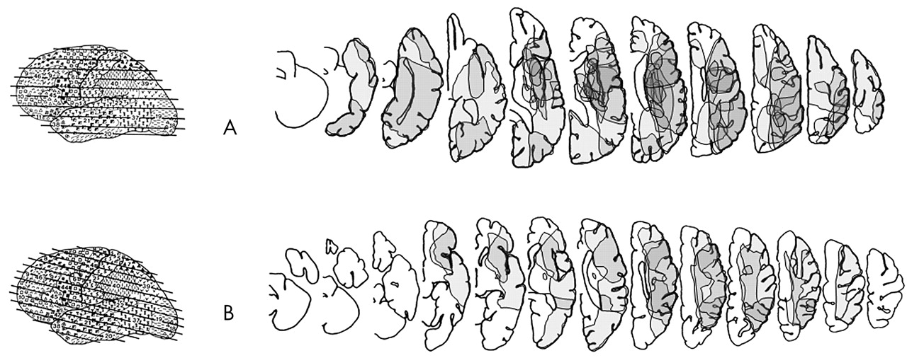
Neglect patients’ lesions. The figure shows graphical reconstructions of lesions (shaded grey areas limited by thin black lines) in the neglect group, as a function of (A) template A18 (15 patients: three MRI and 12 CT scans) and (B) template A20 (four patients: one MRI and three CT scans) from Damasio and Damasio’s atlas. For two neglect patients the CT scan was negative. The degree of overlap among patients’ lesions is represented by the progressive darkening of the grey areas. The scan plane of each template is represented on the left.

No-neglect patients’ lesions. The figure shows graphical reconstructions of lesions and degree of lesion overlap in the no-neglect group, as a function of (A) template A18 (five patients: all CT scans) and (B) template A20 (three patients: all CT scans) from Damasio and Damasio’s atlas. The scan plane of each template is represented on the left. Conventions as in fig 1.
To extract the areas exclusively associated with neglect, computer graphics were used in a two-step procedure of anatomical subtraction between groups’ lesions. First, a neglect minus no-neglect subtraction was performed, that is, areas damaged in no-neglect patients were graphically subtracted from areas damaged in neglect patients, thus revealing areas that were damaged only in neglect patients. This subtractive procedure, by eliminating all the areas whose damage did not induce neglect, leaves areas that are crucial for determining neglect, and possibly areas that are not directly related to neglect, but were damaged along with critical areas in large lesions. Second, to discard the contribution of the large lesions (a few cases), a minimum lesion overlap threshold was chosen. As the remaining areas of overlap for each slice of either template contained two to four overlapping lesions, we selected plots of areas, in each slice of each template, that contained at least three overlapping lesions. This conservative criterion, by reducing the risk of a false positive, increases the reliability and spatial resolution of the procedure (fig 3).

Neglect associated lesions. The figure shows plots of the neglect minus no-neglect graphical subtraction: damaged areas that were exclusively associated with neglect patients, as a function of (A) template A18 and (B) template A20 from Damasio and Damasio’s atlas. The scan plane of each template is represented on the left. Conventions as in fig 1.
Similarly, a neglect-persistence minus neglect-recovery subtraction was performed to locate areas responsible for neglect persistence, that is, the lesions crucially associated with the lack of neglect recovery. In this case, the areas damaged in patients with neglect recovery were subtracted from those damaged in patients without neglect recovery.
RESULTS
Several ANOVAs were performed, and the Newman-Keuls post hoc test was used whenever necessary. To focus on the pattern of spontaneous recovery (table 2), mean values of interactions assessing neglect improvement across sessions will be detailed, in addition to their statistical significance. A Kolmogorov-Smirnov test showed that, in some sessions, the distribution of some RT measures was not normal. As the results of a GLM analysis exactly replicated those obtained with ANOVAs, the latter are reported.
Pattern of spontaneous recovery in acute neglect patients
Non spatial attention
SART
An ANOVA with Group (neglect, no-neglect, healthy) as between-subject factor [F(2,31) = 11.95; p<0.0001] revealed longer RTs at the first session in both neglect (625 ms) and no-neglect (620 ms) patients compared to healthy subjects (414 ms, both p<0.007), patient groups not differing between them. The accuracy in “go” responses was comparable for neglect (66%), no-neglect (67%), and healthy subjects (76%) who, in the “no-go” responses, performed less accurately (45%) than neglect (67%) and no-neglect (78%) groups (both p<0.02). The performance of neglect and no-neglect patients was comparable.
To assess the pattern of recovery, three ANOVAs with Session (first, second, third) as within-subject factor were run. No significant difference was found for RTs in neglect (first: 625 ms, second: 594 ms, third: 596 ms) or no-neglect groups (first: 620 ms, second: 579 ms, third: 589 ms). The accuracy for “go” responses slightly increased in neglect (first: 66%, second: 73%, third: 75%) and no-neglect groups (first: 67%, second: 77%, third: 76%), the “no-go” accuracy being stable in both groups.
Base-dual task
A similar ANOVA, besides showing that RTs of the first session were longer in the dual (1740 ms) than in the base (876 ms) task [F(1,31) = 31.52; p<0.0001], also revealed that neglect (1552 ms) and no-neglect (1586 ms) patients were slower compared with healthy subjects (692 ms, both p<0.05), without differing between them.
To assess neglect recovery, an ANOVA was conducted on RT in both tasks (base, dual) with Session (first, second, third) and Side (left, right) as within-subject factors. In neither analysis was the Group×Session×Side interaction significant. In the base task, neglect patients’ RTs tended to shorten on the left (first: 1422 ms, second: 1055 ms, third: 950 ms) and right sides (first: 927 ms, second: 715 ms, third: 655 ms), and no-neglect patients showed the same trend on the left (first: 1029 ms, second: 871 ms, third: 726 ms) and right sides (first: 834 ms, second: 739 ms, third: 576 ms). On the dual task, neglect patients’ RTs did not ameliorate either on the left (first: 1200 ms, second: 1416 ms, third: 1316 ms) or the right side (first: 894 ms, second: 1021 ms, third: 941 ms). Instead, no-neglect patients tended to improve on the left (first: 1552 ms, second: 693 ms, third: 750 ms) and right sides (first: 1276 ms, second: 659 ms, third: 642 ms).
Spatial attention
An ANOVA with Group (neglect, no-neglect) as between-subject factor, and Side (left, right) and Session (first, second, third) as within-subject factors, was run on accuracy for each test.
Bell cancellation
The Group×Side interaction [F(1,31) = 32.9; p<0.0001] revealed that neglect patients were more accurate on the right (68%) than on the left side (36%, p<0.0001), no difference being present in no-neglect patients (86% and 85%). No-neglect performed better than neglect patients both on the left (86% v 36%, p<0.0001) and the right sides (85% v 68%, p<0.0002). The Group×Session interaction [F(2,62) = 6.6; p<0.003] showed neglect improvement in the second (54%, p<0.003) and third sessions (60%, p<0.0002) compared with the first session (42%). No difference was found in the no-neglect group (first: 87%, second: 84%, third: 86%).
Letter cancellation
Group×Side [F(1,31) = 16.9; p<0.0003] revealed poorer performance for neglect patients only on the left side (80% v 46%, p<0.0002). Neglect patients tended to improve more with time (first: 47%, second: 57%, third: 71%) than no-neglect patients (first: 75%, second: 77%, third: 85%).
Picture scanning
The Group×Side interaction [F(1,31) = 12.8; p<0.001] showed poorer performance for neglect patients only on the left side (52% v 96%, p<0.0001). Group×Session [F(2,62) = 3.2; p<0.05] revealed neglect amelioration from session 1 (57%) to sessions 2 (72%, p<0.02) and 3 (80%, p<0.0009). No-neglect patients’ performance was stable (first: 94%, second: 98%, third: 96%).
Menu reading
The Group×Side interaction [F(1,30) = 10.8; p<0.003] showed that neglect patients were less accurate than no-neglect group on the left (66% v 98%, p<0.0002) but not on the right side (93% v 99%). The nearly significant Group×Session interaction (p<0.09) suggested neglect amelioration (first: 70%, second: 82%, third: 86%). No-neglect patients always performed this task quite accurately (first: 98%, second: 98%, third: 100%).
Line bisection
A similar ANOVA on the mean displacement (mm) showed an almost significant (p<0.09) larger deviation in neglect than in no-neglect patients (17 v 5 mm). Neglect patients also tended to improve across sessions (first: 22 mm, second: 16 mm, third: 14 mm).
Fluff test
Performance was poorer [F(1,31) = 14.2; p<0.0007] in neglect (62%) than in no-neglect patients (94%). The non significant (p<0.1) trend towards amelioration of neglect (first: 54%, second: 59%, third: 75%) was absent in no-neglect patients, who were quite accurate (first: 93%, second: 93%, third: 97%).
Lateralised target
Two ANOVAs were carried out with Group (neglect, no-neglect) as between-subject factor, and Session (first, second, third) and Side (left, right) as within-subject factors. Focussing on the recovery pattern, the non significant interaction Group×Session (p<0.1) suggested an amelioration of RTs in neglect (first: 1232 ms, second: 1034 ms, third: 955 ms) but not in no-neglect patients (first: 890 ms, second: 919 ms, third: 836 ms). Concerning omissions, the Group×Side interaction verged on significance (p<0.08), inasmuch as neglect patients omitted far more items on the left (26%) than the right side (1%), compared to no-neglect patients (2% and 0%). No improvement with time was found in either group.
Lateralised response
Similar ANOVAs revealed that a non significant improvement in RTs was apparent in neglect (first: 2174 ms, second: 1912 ms, third: 1910 ms) and no-neglect groups (first: 2224 ms, second: 1939 ms, third: 1827 ms). Comparable numbers of left- and right-sided omissions were made by patients with (left: 16%, right: 16%) and without neglect (left: 4%, right: 4%). Neglect patients tended to show a slight improvement across sessions (first: 26%, second: 14%, third: 9%), a smaller tendency being manifest in patients without neglect (first: 7%, second: 3%, third: 2%).
Neglect-related disorders
An ANOVA [F(1,27) = 9.7; p<0.004] showed more severe anosognosia in neglect (27%) than no-neglect patients (4%), with no amelioration with time in either group. Similar ANOVAs were run for extinction, with Stimulation (unilateral left, bilateral) as additional within-subject factor.
Visual extinction
The nearly significant Group×Stimulation interaction (p<0.08) suggested more impairment on bilateral (45%) than unilateral stimulation (80%) in neglect compared to no-neglect patients (83% v 96%). To focus on the recovery pattern, a trend towards amelioration was present in neglect patients for unilateral (first: 75%, second: 79%, third: 86%) but not bilateral stimulation (first: 45%, second: 44%, third: 45%). In contrast, no-neglect patients showed little improvement either in unilateral (first: 90%, second: 98, third: 100%) or bilateral stimulation (first: 75%, second: 85%, third: 88%).
Tactile extinction
Neglect patients were impaired in unilateral (67%) and bilateral stimulation (52%), similarly to no-neglect patients (83% v 67%). Also, no improvement with time was found in neglect patients, either in unilateral (first: 68%, second: 69%, third: 64%) or bilateral stimulation (first: 48%, second: 53%, third: 55%), whereas no-neglect patients tended to improve in unilateral (first: 70%, second: 89%, third: 90%) and bilateral stimulation (first: 50%, second: 80%, third: 70%).
Motor abilities
Two ANOVAs were run on movement displacement (MA 90° and 180°).
Wrist and finger
The Group×Movement interaction [F(1,26) = 4.7; p<0.04] showed that no-neglect patients performed the finger extension task (43°) better than neglect patients (18°, p<0.01), the groups not differing in the wrist extension task (24° v 22°). The marginally significant (p<0.06) Group×Movement×Session interaction showed a smaller improvement in the finger extension task in neglect patients (first: 16°, second: 17°, third: 22°) than in no-neglect patients (first: 32°, second: 45°, third: 53°). A comparable amelioration of wrist extension was apparent in neglect (first: 19°, second: 21°, third: 26°) and no-neglect patients (first: 22°, second: 21°, third: 28°).
Elbow and shoulder
No difference was observed between groups in shoulder flexion (75° v 76°) and abduction (68° v 65°), or elbow flexion (77° v 77°). A non significant (p<0.2) improvement was apparent in neglect patients for shoulder flexion (first: 71°, second: 78°, third: 76°) but not shoulder abduction (first: 67°, second: 69°, third: 68°) or elbow flexion (first: 78°, second: 76°, third: 76°). No-neglect patients showed no improvement on shoulder flexion (first: 76°, second: 72°, third: 74°), with little improvement for shoulder abduction (first: 59°, second: 68°, third: 69°) and elbow flexion (first: 72°, second: 80°, third: 79°).
An ANOVA with Hand (left, right) as additional within-subject factor was used for the grip force. Neglect patients’ performance did not change with time for the left (2 kg in all sessions) or the right hand (24 kg in all sessions). The same was true for no-neglect patients for the left (first: 5 kg, second: 5 kg, third: 8 kg) and the right hand (first: 24 kg, second: 26 kg, third: 24 kg).
Chronic follow up
The performance of neglect patients re-examined in the chronic phase (fully detailed in table 3) was similarly analysed; only significant results are reported below.
Follow up investigation of the spontaneous recovery of neglect
Cancellation tasks
In the bell cancellation task, the Session×Side interaction [F(3,21) = 6.9; p<0.002] showed higher accuracy on the left side in the fourth session (78%) than the first (32%, p<0.0002), the second (54%, p<0.002), or the third session (50%, p<0.0006). Right side accuracy was stable. Lower left versus right accuracy was present in the first (32% v 76%, p<0.0002), second (54% v 77%, p<0.002), and third sessions (50% v 86%, p<0.0002), but not in the fourth session (77% v 89%). Similar differences were found for the letter cancellation task (table 3).
Picture scanning
The interaction Session×Side was marginally significant (p<0.056). The left-right difference exceeded 20% in the first (46% v 86%) and second sessions (59% v 94%), decreasing in the third (83% v 98%) and fourth sessions (83 v 98%).
Fluff test
Personal neglect improved [F(3,21) = 3.9; p<0.03] in the fourth (90%) compared with the first session (65%, p<0.04), with an overall trend towards amelioration.
Motor abilities
An improvement was found for elbow and shoulder movements [F(3,18) = 3.8; p<0.03] in session 4 (92°) compared to session 1 (45°, p<0.02). Besides the expected left–right difference in grip force [F(1,5) = 20.5; p<0.006], the improvement with time was not significant (table 3).
Anatomical correlates of acute neglect
The lesions analysis, besides indicating larger damage in neglect (fig 1) than no-neglect (fig 2) patients, also identified (by neglect minus no-neglect subtraction) the regions damaged only in the neglect group (fig 3): F6 (frontal operculum), F7 (prefrontal), F8 (pre-motor and rolandic), and F9 and F10 (paraventricular and supraventricular) in the frontal lobe; P1 and P2 (supramarginal and angular gyrus), P3 and P4 (lateral and medial aspect of SPL), and P5 and P6 (paraventricular and supraventricular regions of SPL) in the parietal lobe.
Anatomical correlates of neglect persistence
The neglect group was divided into recovery and no-recovery groups according to the presence of amelioration in the bell cancellation test, which is most sensitive to improvement with time. When left-side accuracy was analysed (one-tailed Fisher’s test) it was shown that ten patients (43%) improved in the third compared with the first examination, only two of them (9%) showing complete recovery (table 4). When comparing the first with the fourth session, performed by eight neglect patients in the follow up, it was shown that five patients (63%) improved, only one (13%) showing complete recovery.
Spontaneous recovery of extrapersonal neglect
Lesion subtraction (no-recovery minus recovery) identified the areas associated with neglect persistence. In agreement with the previous analysis, these areas largely corresponded to those found to be responsible for neglect, and included: F6, F7, F8, F9, and F10 in the frontal lobe; and P2, P4, P5, and P6 in the parietal lobe.
DISCUSSION
The present study investigated the spontaneous recovery of neglect, particularly focussing on: (a) the spatial/non spatial nature of the attentional deficit, (b) the evolutionary pattern of acute neglect during a 2 week period, (c) the characteristics differentiating neglect recovery from neglect persistence, and (d) the anatomical correlates of neglect presence versus neglect absence, as well as neglect recovery versus neglect persistence. The main results are discussed in the following paragraphs.
Spatial versus non spatial deficits and their evolution with time
The findings did not support the notion that non lateralised attentional deficits can discriminate between neglect and no-neglect patients, although neglect patients were certainly affected by non lateralised attentional deficits compared with age-matched healthy subjects. Both patient groups showed poor sustained and divided attention (SART and base-dual tasks), probably due to impaired functioning of non lateralised attention. At variance with pure tests of sustained attention,71 the SART also involves executive control functions. Thus, the comparably slow performance of neglect and no-neglect patients suggests that non lateralised attention and possibly control functions can contribute to neglect, although not in a selective way, the patient’s deficit being related to the lesion per se without being specifically related to the presence of neglect. These results apparently contrast with those of Buxbaum et al,63 whereby non lateralised attentional deficits correlated with neglect severity, as previously reported by Robertson and colleagues.72 In this respect, since the correlation was not assessed separately in neglect and no-neglect patients, the possibility remains open, to be verified in future studies, that neglect severity is indeed correlated with non lateralised attentional deficits, which are not necessarily responsible for neglect but may nevertheless contribute to the syndrome.73
Considering the evolutionary pattern, neglect patients did not improve in either the SART or the base-dual task. The slight and generalised amelioration in these tasks may reflect practice effects and/or increased arousal. Overall, the results indicated that the two groups of patients did not differ significantly either in terms of sustained attention and executive control, or divided attention. This finding (consistent with Heilman’s hypothesis) suggests that right brain-damaged patients may be hypoaroused following damage of a (right) dominant alertness system.
In contrast, spatial attention deficits were clearly present in neglect. While they were expected in the tasks used here to operationally define neglect (bell, letter cancellation), more ecological tests confirmed the presence of spatial defects (picture scanning, menu reading) also present within personal space (fluff test). Relative to extrapersonal and personal dissociation,15,16 17% of patients with extrapersonal neglect were not affected by personal neglect. Bearing in mind the possible differential sensitivity of individual tasks, this finding confirms that neglect is not an all-or-none phenomenon, but can selectively affect different sectors of space.
Moreover, the spontaneous recovery pattern was consistently characterised by significant, or almost significant, improvements in spatial attention disorders exclusively in neglect patients, both in personal and extrapersonal space. These findings confirm that spatial attention deficits can represent the main characteristic of the neglect syndrome.48,74 Moreover, when compared with no-neglect subjects, neglect patients clearly tended to be slower on left- than right-sided responses, and to omit more left-sided items in the lateralised target task, but not in the lateralised response task in which they did not differ from no-neglect patients. These results provide at least partial support for perceptual/premotor dissociation in neglect,18,19,75,76 confirmed by the selective perceptual impairment present in seven patients.
Overall, the findings strongly support the idea that neglect is mainly characterised by spatial deficits, though non lateralised attentional deficits are also present, without being specifically responsible for the major manifestations of the deficit. Accordingly, not only cancellation tests but most spatial attentional tests were sensitive enough to discriminate between neglect and no-neglect patients.
Neglect-related disorders and their evolution with time
Anosognosia was more severe in neglect than no-neglect patients. It was present in 17 of 23 neglect patients and evolved differentially, not improving as much as spatial deficits during the period of investigation. This favours a dissociation between neglect and anosognosia, although it may have also been influenced by potential differences in the sensitivity of the test used. Similarly, only 12 neglect patients showed visual extinction, and some amelioration for unilateral, but not bilateral, stimulation, the same being true for tactile extinction (present in eight patients). The discordant evolution of neglect (improved) and extinction (not improved) suggests that they may be dissociable disorders with different recovery patterns.
An important issue addressed here was the evolution of neglect-associated motor disabilities. Indeed, contralesional finger movements were more impaired in neglect than no-neglect patients, the latter appearing to gain better distal motor control across sessions compared to neglect patients. No group differences were observed for proximal movements or grip force. This finding confirms the negative impact of visual neglect (improved) on motor recovery (not improved), at least during the acute stage of the illness.
Summarising, only 43% of neglect patients improved spontaneously during the 2 week period. By comparing the accuracy on the bell cancellation task between the first and third sessions, this improvement could be attributed to a less severe category of neglect (table 4). Complete recovery (subclinical neglect) was observed only in 9% of patients.
Greater amelioration of extrapersonal and personal neglect was visible at follow up. Some 63% of patients clearly recovered, although only one of them (13%) reached a subclinical level of severity. Also visual and tactile extinction tended to ameliorate in the chronic stage, whereas anosognosia did not improve. Most interesting, contralesional shoulder and elbow movements improved significantly, while distal movements improved to a lesser extent. The finding of concomitant amelioration of neglect symptoms and contralesional (proximal) motor deficits strongly supports the possibility that motor recovery might be favoured, at least in the chronic stage of the illness, by relieving patients of neglect deficits.
Therefore, spontaneous recovery in the acute phase of the disease is not axiomatic, and, when present, does not allow for complete remission of neglect symptoms in most patients. The amelioration obtained in the chronic phase is more encouraging, but potential implications might be limited by the smaller sample size.
Anatomical correlates of neglect and neglect persistence
Some frontal lobe areas (BA 1–4, 6, 8, 9, 44–46) were found to be crucially associated with neglect. In agreement with the role potentially played by these areas in neglect manifestations is the fact that they contain somatosensory (3, 1, 2), pre-motor (44, 6), and motor (8, 4) maps contributing to the construction of perceived space representation. Lesion of these areas may induce neglect because of the reduced competitive strength of contralateral compared to ipsilateral space representation. In agreement with the contribution provided by sensorimotor information for space representation, several studies showed that contralesional motor and proprioceptive stimulation ameliorates neglect.77–79
Some parietal areas (BA 5, 7, 39, 40) were also critically associated with neglect, as previously reported.2,46 Thus, the present study does not support a privileged role being played by temporal lesions in the genesis of acute3 or chronic neglect.48,80 Indeed, temporal structures were not crucially associated with neglect. The present anatomical results could have been limited by the relatively small number of patients investigated, and by the relatively coarse spatial resolution allowed by a standard procedure based on CT/MRI scans acquired for clinical purposes with different scan planes. However, the lack of correspondence between temporal lesions and neglect has been recently confirmed in a large group study,63 as well as by Mort and colleagues,49 who used a high resolution technique of brain lesion mapping.
Finally, the no-recovery minus recovery comparison revealed that areas associated with neglect persistence (BA 1–9, 39, 44–46) largely corresponded to those found to induce neglect. The similarity of lesion location between transitory and persistent neglect observed in the present investigation is consistent with the findings of a longitudinal study61 and other reports identifying in a parieto-frontal circuit the most likely candidate responsible for neglect.81 It is interesting to note that, in agreement with the results recently reported by Mort and colleagues,49 the angular gyrus (BA 39) was found to be involved both in the acute genesis (neglect minus no-neglect) and in the maintenance (no-recovery minus recovery) of the neglect syndrome.
Therefore, the present findings indicate that somatosensory, premotor, and motor areas play an important role in the genesis and maintenance of neglect, as their integrity may be necessary for neglect recovery.82 Further longitudinal studies of the recovery of visuospatial neglect and its anatomical correlates would hopefully clarify the relationships between the location of brain damage and the recovery of visuospatial neglect.
Acknowledgments
We wish to thank all the subjects for their collaboration. We are grateful to F Pavani and T Ro for helpful discussions and to R Bolzani and MG Benassi for assisting with statistical analyses.
REFERENCES
Footnotes
-
↵* Current address: University of Houston, Houston, TX, USA
-
This work was supported by a grant to LB from the James S McDonnell Foundation.
-
Competing interests: none declared