Article Text

Abstract
Objectives Recently, IgG autoantibodies against different paranodal proteins have been detected and this has led to important advances in the management of inflammatory neuropathies. In contrast, not much is known on IgM autoantibodies against paranodal proteins.
Methods In the present study, we screened a large cohort of patients (n=140) with inflammatory neuropathies for IgM autoantibodies against neurofascin-155, neurofascin-186 or contactin-1.
Results IgM autoantibodies against neurofascin-155 were detected by ELISA in five patients, four with inflammatory demyelinating polyradiculoneuropathy (CIDP) and one with Guillain-Barré syndrome (GBS), and were confirmed by ELISA-based preabsorption experiments and Western blot. Titres ranged from 1:100 to 1:400. We did not detect IgM anti-neurofascin-186 or anti-contactin-1 antibodies in this cohort. All patients presented with distally accentuated tetraparesis and hypesthesia. Remarkably, tremor was present in three of the patients with CIDP and occurred in the patients with GBS after the acute phase of disease. Nerve conduction studies revealed prolonged distal motor latencies and F wave latencies. Nerve biopsies showed signs of secondary axonal damage in three of the patients, demyelinating features in one patient. Teased fibre preparations did not demonstrate paranodal damage.
Conclusion In summary, IgM neurofascin-155 autoantibodies may be worth testing in patients with inflammatory neuropathies. Their pathogenic role needs to be determined in future experiments.
Statistics from Altmetric.com
Introduction
IgG-autoantibodies against the paranodal proteins contactin-1, caspr and neurofascin-155 have recently been described in patients with inflammatory neuropathies and are associated with a severe predominantly motor phenotype, subacute onset and often poor response to intravenous immunoglobulins (IVIG).1–6 Patients with anti-neurofascin-155 antibodies often suffer from a debilitating tremor that was suggested to be of cerebellar origin.2 IgG4 was the most frequent subclass of paranodal autoantibodies, but other subclasses of autoantibodies also occur.1 2 5 7 There is evidence that the subclass of autoantibodies may be a predictor of complement activation and response to treatment with IVIG.8 A patient with a combination of IgM, IgA and IgG3 anti-neurofascin-155 autoantibodies has been reported,4 but subsequent studies focused on the detection of IgG autoantibodies. Only recently a cohort of patients with inflammatory and non-inflammatory neuropathies was published that had been screened for anti-neurofascin IgM and IgG by cell-based assays.7 These authors found IgM autoantibodies in 8% of patients with inflammatory neuropathy, but also in 5% of patients with idiopathic neuropathy. In contrast to IgG autoantibodies against paranodal proteins, the clinical relevance and the clinical picture of IgM autoantibodies is unknown. In the present study, we aimed to determine the prevalence of anti-neurofascin-155, anti-neurofascin-186 and anti-contactin-1 IgM in a cohort of patients with inflammatory neuropathies who were tested negative for IgG autoantibodies and to characterise their clinical phenotype.
Methods
Patients
Between 2011 and 2017, 102 patients who attended the Department of Neurology of the University Hospital Würzburg for diagnostic work-up or therapy of neuropathies and two patients from the University Hospital Jena, Department of Neurology, were prospectively included into the study. Seventy-seven of these patients fulfilled the INCAT/EFNS criteria of chronic inflammatory demyelinating polyradiculoneuropathy (CIDP),9 10 29 did not fulfil these diagnostic criteria but were categorised as clinically suspected CIDP. Additionally, 36 samples of plasma exchange material of patients with Guillain-Barré syndrome (GBS) or CIDP that were stored in our department and 82 healthy controls who had partly been recruited for a former study were included.5 Sera of 45 patients with non-inflammatory neuropathies and 16 patients with other autoimmune neurological diseases (myasthenia gravis, autoimmune encephalitis, stiff person syndrome or multiple sclerosis) served as disease controls. No anti-neurofascin-155/-186 or anti-contactin-1 autoantibodies of the IgG type were detected by ELISA in any patient and all sera were additionally screened for IgG autoantibodies against paranodal proteins by binding assays with murine teased fibres as previously described4 5 11 and did not show any paranodal or nodal binding. Some of the patients were included in former studies on IgG autoantibodies against paranodal proteins.5 6 All patients and controls gave informed consent to participate.
ELISA and complement binding assay (CBA)
Maxisorb 96-well plates (Thermo Fisher Scientific, Waltham, Massachusetts, USA) were coated with human contactin-1 (2 µg/mL, Sino Biological, Beijing, China), human neurofascin-155 or neurofascin-186 (5 µg/mL)4 and blocked with blocking solution (PBS, 3.33 %BSA, 0.05% Tween20). All wells were incubated with patient sera diluted 1:100, anti-contactin-1 (mouse monoclonal, Abcam, Cambridge, UK, 1:200) or anti-pan-neurofascin (chicken polyclonal, R&D Systems, Minneapolis, Minnesota, USA, 1:1000) as controls. Peroxidase-conjugated secondary antibodies (anti-human IgM, Dako, Glostrup, Denmark, for patient material, 1:7000 or anti-mouse IgG/anti-chicken IgY, 1:10 000, for the control antibodies) were used and optical density was measured at 450 nm with Multiscan EX ELISA reader (Thermo Fisher Scientific). Normal cut-off values were based on the measurements of normal controls and were set at five standard evaluations above the mean of all normal controls.5 11 Titres of IgM anti-neurofascin-155 autoantibodies were determined by diluting all positive sera from 1:100 to 1:1200 and the highest dilution above the cut-off value was taken. The number of anti-neurofascin-155 IgM-positive patients and normal and disease controls was compared using two-sided Fisher’s exact test.
For the CBA, Maxisorb 96-well-plates were coated, blocked and incubated with patient and control sera (1:20) as described above. Between incubations, six washing steps with PBS+0.05% Tween 20 were performed. Serum of a patient with neurofascin-155 IgG autoantibodies (titre 1:6000) was used as a positive control. The wells were then incubated with C1q (10 µg/mL, Sigma Aldrich, St. Louis, Missouri, USA) for 2 hours at room temperature, following incubation with peroxidase-conjugated anti-C1q (LifeSpan Biosciences, Seattle, Washington, USA, 1:200) for 30 min. Optical density was measured with the ELISA reader as described above.
Preabsorption experiments
Preabsorption was performed by serial incubation of the sera of all anti-neurofascin-155-positive patients and a control (1:100 in blocking solution) on an ELISA plate coated with neurofascin-155 (as described above) or contactin-1 (5 µg/mL). Doublets of all diluted sera were serially incubated in six steps on coated wells for 1 hour each. Afterwards, immunoreactivity to neurofascin-155 was tested by ELISA as described above. Sera of all patients that were not preincubated were run as controls.
Western blot
Neurofascin-155 protein (6 µg/lane)4 was loaded onto polyacrylamide gels for electrophoresis. Protein fractions were blotted onto nitrocellulose membranes and incubated with patient sera and anti-neurofascin-155 (1:200, rabbit, polyclonal, Abcam) as a control. Peroxidase-conjugated anti-human IgM (1:5000, Dako, Glostrup, Denmark) was used as a secondary antibody for patient material, anti-rabbit IgG for the controls. Protein bands were detected using ECL Plus Detection Kit (Perkin Elmer, Waltham, Massachusetts, USA).
Binding assays on neurofascin-155 transfected human embryonic kidney 293 (HEK293) cells and murine teased fibres
Binding assays on HEK293 cells transfected with a plasmid of human neurofascin-155 and on murine teased fibres were performed as previously described,4 11 using Cy3-conjugated anti-human IgM as a secondary antibody (Dianova, Hamburg, Germany, 1:100).
Histological procedures
Nerve and skin biopsies were taken for diagnostic purposes in some of the patients and were processed according to standard procedures.12 13 Intraepidermal nerve fibre density was calculated following established counting rules.13 Of some nerve biopsies, one part was used for the preparations of teased nerve fibres by gently teasing the fibres prefixed with paraformaldehyde 4% on a slide. Immunofluorescence double-staining with antibodies against myelin basic protein (GeneTex, 1:200) and Caspr (Abcam, 1:100), pan-neurofascin (Abcam, 1:400) and pan-sodium-channel (Sigma Aldrich, 1:100) was then performed as previously described.6 Skin biopsy sections were stained with the same combinations of antibodies for longitudinal assessment of dermal myelinated fibres according to protocols of a former study.14
Results
Patients and samples
Demographic data of all patients are summarised in table 1. Of the patients seen in our Department and at the University Hospital of Jena, 77 were diagnosed as CIDP based on the INCAT and/or EFNS criteria,10 15 34 suffered from GBS, all fulfilling the Brighton criteria.16 Twenty-nine patients with clinically suspected CIDP who did not fulfil the INCAT/EFNS criteria were also included. Monoclonal gammopathy was found in 11 patients (3 IgG kappa, 3 IgM kappa, 2 IgG lambda, 2 IgM lambda, 1 IgA kappa) by routine immunofixation assays. Demographic data are given in table 1.
Summary of the demographic data of all study participants
Detection of five patients with anti-neurofascin-155 IgM by ELISA
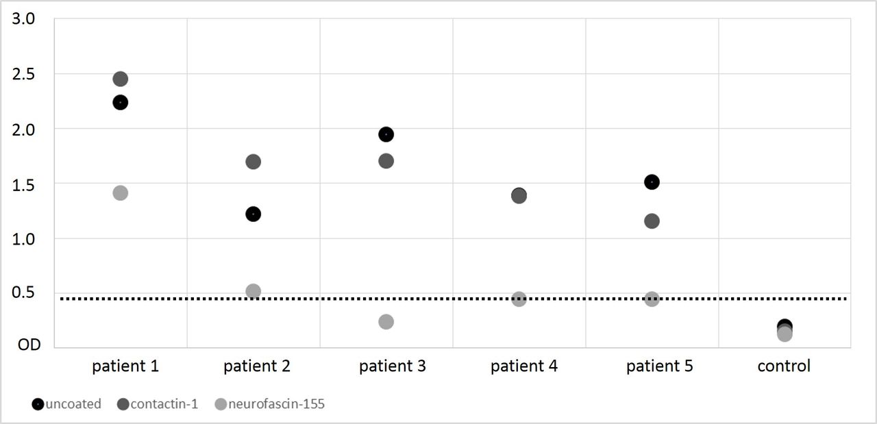
Of the 140 patient samples that were tested by ELISA, immunoreactivity of IgM against neurofascin-155 clearly above the cut-off value was detectable in five patients, not in any control (figure 1) and thus was more often found in patients than controls (p=0.029). We did not detect any immunoreactivity of patient IgM against neurofascin-186 or contactin-1 (data not shown). Titres of IgM anti-neurofascin-155 autoantibodies ranged from 1:100 to 1:400 (see table 2). Binding to neurofascin-155 transfected HEK293 cells as well as murine teased fibres, however, was similar to the background staining of healthy controls. For comparison of the sensitivity of ELISA and the binding assay with neurofascin-155 transfected HEK cells, serum of a patient with anti-neurofascin-155 IgG autoantibodies was tested by both assays in parallel and anti-neurofascin-155 IgG autoantibodies were detectable up to a dilution of 1:6000 by ELISA and up to 1:2000 by binding to neurofascin-155 transfected HEK293 cells, indicating a higher sensitivity of ELISA compared with cell-based binding assays.

Optical density (y-axis) of the samples of the different patient and control groups (x-axis) as measured by ELISA for anti-NF155 reactivity. The dashed line marks the cut-off value of the normal range, which was set five SD above the mean of the healthy controls. Five patients show reactivity above the normal range. CIDP, chronic inflammatory demyelinating polyradiculoneuropathy; GBS, Guillain-Barré syndrome; NP, neuropathy.
Demographic, immunological, clinical and therapeutic data of anti-neurofascin-155-IgM-positive patients (in the order of antibody titres)
We did not detect any C1q binding induced by neurofascin-155 binding in any of the five patients who were positive for anti-neurofascin-155 IgM by ELISA but could detect C1q deposition in a patient with high titre anti-neurofascin-155 IgG autoantibodies who served as a positive control.
Confirmation of anti-neurofascin-155 IgM by preabsorption experiments
Preabsorption of all anti-neurofascin-155-IgM-positive sera as detected by ELISA and a control sample was performed on neurofascin-155 coated ELISA wells. Contactin-1 coated wells served as controls. Preabsorption on neurofascin-155 coated wells resulted in a marked decrease of immunoreactivity in the subsequent ELISA (ratio neurofascin-155/uncoated: median 0.32, range 0.12–0.62), supporting the specificity of anti-neurofascin-155-IgM autoantibodies in these patients. No changes were detectable after preincubation on contactin-1 coated wells (ratio contactin/uncoated: median 0.99, range 0.76–1.39) (figure 2).
Optical density (y-axis) of the samples of the neurofascin-155-IgM-positive patients (x-axis) as measured in the ELISA after preincubation of the sera with contactin-1-coated (dark grey) or neurofascin-155-coated (light grey) wells compared with native sera (black). The dashed line marks the cut-off value of the normal range. Preincubation with neurofascin-155, but not preincubation with contactin-1-coated wells, led to a decrease of optical density in the ELISA.
Detection of anti-neurofascin-155 IgM by Western blot
To evaluate the specificity of anti-neurofascin-155 IgM autoantibodies detected by ELISA, Western blot was performed with all positive sera. Immunoreactivity of IgM against neurofascin-155 was detectable in four of the five patients who were positively tested by ELISA. No immunoreactivity was detectable in six normal controls (figure 3).

Western blot of the samples of patients 1–5, two normal controls (right lanes, all incubated with anti-human IgM), a commercial anti-neurofascin control antibody (chicken polyclonal, R&D Systems) as positive control (left lane, anti-rabbit IgG) and negative controls (anti-rabbit IgG and anti-human IgM, left lanes). Sera of patients 1, 2, 3 and 4 show strong immunoreactivity of IgM to neurofascin-155, comparable to the positive control whereas patient 5 and two normal controls only show some background staining, but not a positive band at 155 kDa.
Clinical picture of patients with anti-neurofascin-155 IgM
Clinical data of the patients are summarised in table 2 and detailed data of nerve conduction studies (NCS) in the online supplementary table 1. Patient 4 was diagnosed as GBS with a typical monophasic course of disease. The four other patients were diagnosed as chronic inflammatory neuropathy, two fulfilling the INCAT and/or EFNS criteria of CIDP.9 10 All patients suffered from progressive distal more than proximal tetraparesis and distal hypesthesia and reported neuropathic pain. Two of the patients had an acute onset of disease with a relapsing-remitting course, two showed a slowly progressive course of disease. Remarkably, tremor was present in three of the four patients with CIDP (action tremor or postural tremor) and occurred in the patient with GBS after the acute phase of disease. The tremor was classified as disturbing by all patients and even disabling in one. Monoclonal gammopathy was excluded in all five patients. CSF protein levels were elevated in patients 1–3 and correlated with autoantibody titres (Spearman’s correlation test, R=0.95, p<0.014), possibly reflecting the extent of radiculitic involvement. Neuropathic pain was reported by four patients. NCS revealed a prolonged distal motor latency (DML) and prolonged F-wave latencies or absent F-waves, only moderate reduction of nerve conduction velocity (NCV) and compound muscle action potentials (CMAP) and sensory nerve action potential (SNAP) in all patients (online supplementary table 1). Spontaneous activity was detectable by EMG in four of five patients, indicating axonal damage. Response to treatment differed between patients with good response to IVIG in four patients, poor response to IVIG but good response to plasma exchange and rituximab in patient 1.
Supplemental material
Follow-up sera were available in three patients (patients 1, 4, 5). In patient 1, neurofascin-155 IgM autoantibodies were detectable at the first admission to our hospital, 5 months after onset of disease, as described above. He then suffered from disabling tetraparesis, distal hypesthesia, tremor and neuropathic pain. After treatment with rituximab, symptoms almost completely remitted and at a control visit 10 months after study inclusion, the patient only complained of mild hypesthesia of the toes. Neurological examination confirmed full strength and only revealed a mild postural tremor of the hands, reduced vibration sense at the hallux and hypesthesia of the toes. NCS also showed marked improvement, with only slightly increased DML and F wave latencies and mild reduction of CMAP and NCV. The patient was able to work full-time again without limitations. No neurofascin-155 IgM was detectable at this time-point.
In patient 4, who was diagnosed with GBS, sera were obtained at the date of admission to hospital (1.5 weeks after onset), 1 week and 2 weeks after admission and at a control visit 1 year after recovery. Neurofascin-155 IgM was detectable at the date of admission and 1 week later, but was not detectable 2 weeks after admission (after treatment with IVIG and PE) and after recovery. In patient 5, who responded moderately to treatment with IVIG, sera were obtained in 2013, 2014 and 2017 and anti-neurofascin-155-IgM was detectable at all time points. Symptoms showed an undulating course of disease during this time period.
Histopathological characterisation of patients with anti-neurofascin-155 IgM
We analysed nerve biopsies of all four patients with CIDP and skin biopsies of two patients with CIDP and of the patient with GBS. Nerve biopsies of three of the patients showed the histological picture of axonal damage with loss of axons, acute degeneration and clusters of regenerating fibres (figure 4). No demyelinating features like thinly myelinated fibres or onion bulbs were detectable. In patient 3, onion bulbs and thinly myelinated fibres as well as signs of axonal damage were found (figure 4). Increase of T cells around epineural vessels were detectable in patients 1 and 3 (figure 5). Immunohistochemistry with anti-Caspr, anti-neurofascin and anti-pan-sodium channels of freshly teased fibres was available in patients 1 and 5. No dispersion of paranodal or nodal proteins and no elongated nodes were found in these two patients. Skin biopsies were done in three patients and showed a normal intraepidermal nerve fibre density in patient five and a slight reduction in patients 1 and 4. No abnormalities of paranodal or nodal proteins were found in dermal myelinated fibres.
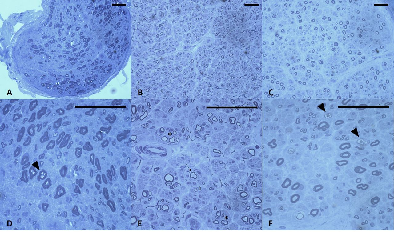
Photomicrographs of semithin sections of nerve biopsies of patient 1 (A,D), patient 3 (B,E) and patients 5 (C,F). Loss of axons was found in all three nerve biopsies (A–C). Nerve biopsies of patients 1 and 5 were classified as axonal neuropathy due to axonal loss and acute degenerating fibres (D,F; arrow heads). The nerve biopsy of patient 3 (B,E) was classified as mixed neuropathy due to axonal loss, regenerating clusters (asterisk) and onion bulbs (arrow). Bar=100 µm.

Photomicrographs of immunohistochemical stains with anti-Leu4, directed against T cells. Accumulation of T cells around epineural vessels was found in patient 1 (A,B) and patient 3 (C,D). Bar=100 µm.
Discussion
In the present study, we give the first detailed work-up of patients with IgM autoantibodies against neurofascin-155. Whereas IgG autoantibodies to neurofascin-155 have been reported in several studies and are supposed to cause a distinct clinical phenotype,2 17 18 IgM neurofascin-155 autoantibodies have so far only been described in a few patients and some of them also tested positive for IgG anti-neurofascin.4 7
Autoantibodies were detectable by ELISA and were confirmed by preabsorption experiments and by Western blot in four patients, but not by binding assays with neurofascin-155 transfected HEK293 cells or murine teased fibres. Most likely, the latter methods were not sensitive enough to detect the low titres of autoantibodies as indicated by a higher sensitivity of ELISA when comparing with cell based binding assays using serum of a patients with high titres of anti-neurofascin-155 IgG.
C1q deposition was not detectable by CBA although IgM antibodies are supposed to bind complement. Complement activation induced by IgM autoantibodies was recently detected in patients with GM1 IgM but only at titres of more than 1:40019 and was shown to correlate with anti-GM1 IgM titres.20 21 Thus, it is not surprising that CBA did not reveal any complement deposition in our sera with only low titre of IgM autoantibodies. Complement deposition was shown for IgG autoantibodies against paranodal proteins, but at much higher autoantibody titres.6 8 22
The patients with IgM neurofascin-155 identified in our study share some features that have also been described in patients with IgG anti-neurofascin-155: tremor was present in four of five patients with IgM neurofascin-155. Although the tremor was less disabling in these patients than in the patients with IgG-autoantibodies, it is of interest that even the patient with GBS developed tremor after the acute phase of disease when the severe tetraparesis had improved, possibly as a residual symptom. There is evidence that tremor in neurofascin-155 IgG-positive patients may be caused by autoantibody binding to cerebellar structures;2 however, we could not detect cerebellar binding in our patients (data not shown), possibly due to the much lower titre of IgM anti-neurofascin-155 compared with IgG titres in former studies. Neuropathic pain is not a typical feature of neurofascin-155-IgG-associated neuropathy but was reported by four of our patients. We can only speculate on the underlying pathogenesis: inflammation induced by IgM autoantibodies may be an explanation as well as damage of small fibres that might correspond to the slight reduction of intraepidermal nerve fibre density in two patients.
NCS of patients with anti-neurofascin-155-IgG are generally characterised by prolonged DML, conduction blocks, decreased CMAP/SNAP and moderate reduction of NCV, features that are in line with the idea of a paranodal or nodal dysfunction rather than demyelination. Indeed, the term paranodopathy/nodopathy was recently suggested to better describe the pathogenesis of neuropathies that target the nodal region.23 24 NCS of our patients did not show any conduction blocks, but prolonged DML and loss of F waves that might be consistent with proximal conduction failure or distal slowing. However, no nodal or paranodal damage was detectable in teased nerve fibres or dermal myelinated fibres that allow immunohistochemical analysis of nodal architecture. In contrast, severe damage to the distribution of paranodal and nodal proteins and ultrastructural architecture was described in patients with IgG autoantibodies against paranodal proteins.5 17 25 26 This argues against a destructive effect of IgM autoantibodies at the nodes, in contrast to the IgG autoantibodies. A pathogenic effect of anti-neurofascin IgM is supported by a recent study that reported enhancement and prolongation of experimental autoimmune neuritis in rats after injection of anti-neurofascin IgM, but not by injection of an isotype control.4 The different subclasses may also explain differences in therapeutic response: our patients mostly showed good response to treatment with IVIG that is supposed to decrease complement deposition,8 whereas patients with anti-neurofascin-155 IgG4 have been shown to be unresponsive to IVIG.2 Almost complete recovery of symptoms was associated with complete decline of autoantibodies in patients 1 and 4 after treatment with rituximab and PE respectively and supports the idea of these autoantibodies to be disease-associated. Similar recovery after treatment with rituximab were reported in patients with paranodal autoantibodies of the IgG type.5 6 27
In contrast to a recent study also describing anti-neurofascin-155 IgM,7 we did not detect any IgM autoantibodies in patients with non-inflammatory neuropathies and thus found a significant difference of autoantibody frequency between inflammatory neuropathies and controls. This supports the idea of a pathogenic or at least disease-associated role of IgM autoantibodies.
In summary, our study provides a detailed description on anti-neurofascin-155 IgM autoantibodies in patients with the clinical picture of CIDP and GBS. Our study was not designed to prove pathogenicity of these autoantibodies. The titres are much lower than titres that were described for IgG autoantibodies against paranodal proteins,2 4 5 and although some clinical features indicate a common phenotype, we cannot give evidence of nodal or paranodal destruction that would support the pathogenicity of IgM autoantibodies in these patients. Further studies are needed to clarify if IgM autoantibodies present an epiphenomenon that occurs in a subgroup of autoimmune neuropathies or play a pathogenic role in these patients. However, IgM anti-neurofascin-155 autoantibodies may be a further diagnostic marker to identify patients with inflammatory neuropathies, even detectable in two patients who did not fulfil the diagnostic criteria of CIDP.
Acknowledgments
The authors thank Barbara Reuter and Susanne Hellmig for expert technical assistance.
References
Footnotes
Contributors KD: study conception and design, analysis and interpretation of data, writing of the manuscript. HS: performance of experiments, data collection and analysis, revision of the manuscript. LA: performance of experiments, data collection and analysis, revision of the manuscript. JG: data collection and analysis. JKMN: study conception. EM: study conception, interpretation of data, revision of the manuscript. CS: study conception and design, interpretation of data, revision of the manuscript.
Funding The study was funded by a grant of Kedrion International GmbH (KD, CS) and the German KKNMS (Kompetenznetz Multiple Sklerose) (EM).
Competing interests KD received personal fees from Baxter/Baxalta and Grifols. EM received personal fees from Roche, Novartis and Genzyme and grants outside the submitted work from Novartis and Genzyme. CS received personal fees from Air Liquide, Alnylam, Astellas, Baxalta, CSL Behring, Genzyme, Grifols, Pfizer, UCB and Kedrion.
Patient consent Parental/guardian consent obtained.
Ethics approval Ethic's committee of the University of Würzburg.
Provenance and peer review Not commissioned; externally peer reviewed.
Linked Articles
- Editorial commentary