Article Text

Statistics from Altmetric.com
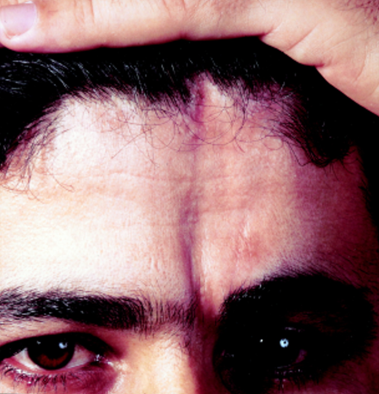
A 28 year old man of Turkish descent had a five year history of monthly nocturnal epileptic attacks. Physical examination disclosed a linear, atrophic, brownish skin area on the left forehead just lateral to the midline. The lesion extended from the left superior orbital ridge into the scalp and was associated with hair loss. There was no facial hemiatrophy (figure A) Neurological examination disclosed no abnormalities. EEG was normal. Brain CT showed small intracranial calcifications in the left temporal lobe and loss of subcutaneous tissue at the site of the lesion (arrows) without involvement of skull bone (figure B). Brain MRI was normal.

If, as in our patient, the history is unclear, this lesion can be mistaken for a scar. However, it is typical of linear scleroderma en coup de sabre, a rare form of localised scleroderma. This is often associated with neurological symptoms, especially epilepsy. Intracranial calcifications or white matter abnormalities can occur. Because the pathogenesis is still unknown, it remains a matter of debate whether cerebral involvement in linear scleroderma en coup de sabre is a consequence of an inflammatory process or constitutes a neurocutaneous syndrome.1 2