Article Text

Statistics from Altmetric.com
Headache is a common, although underemphasised, complaint of lateral medullary infarction. More than half of patients with posteroinferior cerebellar artery (PICA) infarcts develop headache. Fisher found headache in 22 out of 41 (54%) patients with lateral medullary infarction and, more recently, Kuwabara and Hirayama reported headache in 26 out of 34 (76%) patients with Wallenberg's syndrome.1 2 Less than 5% of patients with PICA infarcts, however, develop periocular, hemicranial headache and although sympathetic dysfunction in the form of Horner's syndrome, is a well known manifestation of lateral medullary infarction,1 2 signs of parasympathetic overactivity, such as lacrimation, eye injection, and nasal congestion, have never been described in lateral medullary infarction. Here we report on a patient with a lateral medullary infarction who developed anterior hemicranial pain accompanied by severe and persistent autonomic parasympathetic activation.
This heavy smoking 37 year old man came to our hospital due to acute vertigo, lack of coordination of his left limbs, numbness in his right limbs, dysarthria, and dysphagia. An angiographic study performed in another hospital 3 months earlier due to intermittent claudication and decrease in left radial pulse had disclosed atherosclerotic changes in the lower limbs and in the left subclavian arteries. General examination showed reduced pulses in the left radial and right dorsalis pedis arteries. Abnormal signs at neurological examination included hiccup, nystagmus, left Horner's syndrome, and facial hypoesthesia, left velopalatine weakness, right hemicorporal hypotonia, and ataxia as well as impaired sensation over the right limbs. Cranial MRI showed an acute infarction restricted to the left PICA territory (fig 1), and an occlusion of the left vertebral artery, with no sign of dissection, was seen on angiography. He was treated with heparin and then oral warfarin.

T2 weighted MRI showing an infarct in the left PICA territory.
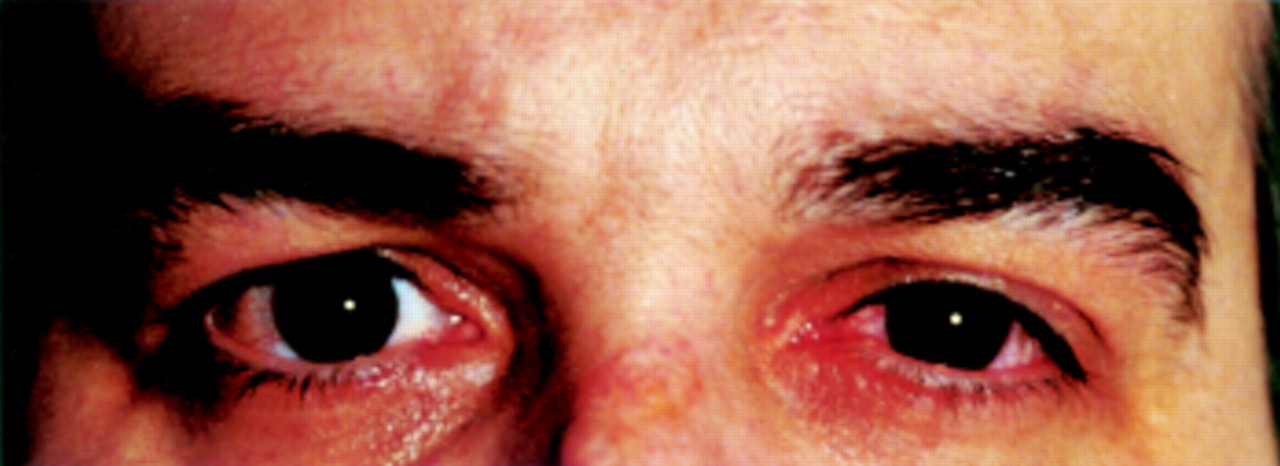
From the beginning of his clinical picture this patient complained of two clearly differentiated head pains. The first one was located in the right occiput and progressively disappeared during the first week after the acute stroke. The second was a very disturbing, continuous headache located anteriorly, mainly in the left retro-orbital and temporal region. This pain was described as moderate to severe, steady, or boring and constantly accompanied by ipsilateral conjunctival injection, lacrimation, and nostril blockage or rhinorrhoea (fig 2). The patient also had one or two dramatic daily exacerbations of unbearable pain intensity together with an increase in autonomic symptoms and signs and lasting about 2–4 hours. The pain did not significantly respond to either oral or intravenous analgesics (paracetamol, aspirin, metimazol, and NSAID). Verapamil, 240 mg daily, plus sodium naproxen, 1100 mg daily, slightly reduced the pain for 2 or 3 weeks. Medications containing ergotamine, methysergide, and agonists of the 5-HT1B/D receptors were not prescribed. The pain remained unchanged for 3 months, then it began to progressively improve and disappeared 6 months after the stroke.

Close up picture of the patient's eyes showing left palpebral ptosis and conjunctival injection.
To the best of our knowledge, this is the first reported patient with a lateral medullary infarction with unilateral anterior headache, sympathetic dysfunction, and parasympathetic autonomic activation, all this resembling a “continuous” cluster-like headache syndrome. The pathophysiology of trigeminal autonomic cephalalgias, including cluster syndrome, is largely unknown.3 It has been proposed, on anatomical grounds, that an inflammatory process in the cavernous sinus, as a point of intersection of the first division of the trigeminal nerve and the cranial sympathetic and cranial parasympathetic nerves, would be the pathophysiological explanation for these headaches.4
As commented on, headache is a frequent complaint in lateral medullary infarction. The posterior is the most common location for headache, 65% in the series of Kuwabara and Hirayama and 49% in Fisher's series,1 2 which concurs with Wolff's finding than on stimulating the vertebral artery the pain is referred to an occipital-suboccipital-nuchal area.5 This posterior pain was also reported by our patient during the first week, probably being related to the thrombus formation in the vertebral artery. The most important pain experienced by our patient, however, was a moderate to severe, unilateral, retro-ocular headache. Anterior pain affecting the eye region is much less frequent in lateral medullary infarction, but was already considered by Fisher as typical of this syndrome and probably resulting from a lesion in the nucleus of the descending root of the trigeminal nerve, as anterior pain cannot be induced on stimulating the vertebral artery and as this anterior headache is usually succeeded by numbness.1 The central medullary lesion also accounts for the autonomic symptoms and signs seen in this patient. Sympathetic dysfunction (Horner's syndrome) is a well-known sign of Wallenberg's syndrome due to damage of the sympathetic tract within the PICA infarction and it seems logical to try to explain the parasympathetic activation as secondary to irritation of the adjacent superior salivatory nucleus by the infarct area or to an imbalance between the two autonomic systems.
In conclusion, this patient shows for the first time that all the semeiology typical of cluster and other trigeminal-autonomic headaches can be secondary to a pure central lesion, located in the lateral medulla, this supporting the contention that flow changes occasionally found in cluster headache cases in the cavernous sinus6 do not generate the disorder, but are in fact a consequence to the pain.7 8 This agrees with the proposal that the activation of the cavernous sinus region does not relate specifically to cluster headache, but it is a trigeminovascular autonomic reflex to first division pain. In fact, the only difference between our case and typical cluster headache—the periodicity of the pain attacks—is easily explained by the distinct central generators giving rise to trigeminovascular system activation— the hypothalamus for idiopathic cluster headache9 and the established lesion of the key structure of this system, the trigeminal nucleus caudalis, in this case.